In 98% of the cases malignant tumours of the testis are germ cell tumours (GCT), divided into pure seminoma (Seminoma) and non-seminomatous GCT (NSGCT) in 60 and 40% of the cases [1].
Germ cell tumours (GCT) are rare, with 2,769 new cases in France in 2018 compared to 1,319 in 1990 [1]. An increase in incidence was reported in France (+21.3% between 2000 and 2014) [2] and worldwide, for example, in Canada where it rose from 3.77/100,000 in 1971 to 7.06/100,000 in 2015 [3]. A further estimated increase of 28% is expected by 2025 [4].
The peak incidence is between 30 and 34 [5] or even 35 years of [2] age, with seminomas appearing later, at around 39 years of age on average.
There is a consensus on certain risk factors for GCT [6]: testicular dysgenesis syndrome (cryptorchidism, hypospadias, hypo- or infertility), personal or family history (1st degree) of GCT and testicular atrophy (<12 ml). Others are being discussed, such as heavy cannabis use [7], pesticides, organic solvents and morphometrics. One study reported an association (OR 3.32, p=0.04) between GCT and first-degree maternal family history of ovarian cancer [8], but it is not consistent with a Swedish registry study [9].
Isolated testicular microlithiasis should not be considered as a risk factor, but its association with known risk factors may call for semi-annual or annual ultrasound surveillance and histological comparison in case of focal lesions [10].
|
|
Diagnosis and pre-therapeutic assessment |
|
| Symptoms and clinical context |
In general, GCT is often diagnosed as a result of local symptoms: palpation of a hard, asymptomatic scrotal mass, sometimes during a painful episode. There is no affinity to a particular side. Bilateral synchronous involvement is exceptional and the disease is localised in 60 – 70% of the cases.
In less than 5% of the cases, the diagnosis will be suggested as a result of regional or general symptoms: abdominal pain or mass, respiratory distress, gynaecomastia, Virchow’s node [11].
|
| Paraclinical examinations |
The determination of 3 serum markers is systematically recommended before and after orchiectomy, for their diagnostic and prognostic value respectively. Negative markers do not eliminate the diagnosis. It is their postoperative value that is the basis for prognostic classifications.
The interpretation of their kinetics requires knowledge of their half-life:
• | Alpha-fetoprotein (AFP) is mainly produced by endodermal sinus tumours and embryonic carcinoma. It is not secreted by seminoma or choriocarcinoma. Its half-life is 5–7 days. It is non-specific and can be found in digestive oncology (hepatocellular carcinoma, and pancreatic and stomach cancers) and in certain non-tumour liver diseases.
|
• | Total human chorionic gonadotropin (hCGt) is routinely secreted by choriocarcinoma but can be found in some embryonic carcinomas and in 10–20% of seminomas. Its half-life is 24–36 hours. The levels may appear high in case of elevation of the ß subunit of LH or pituitary production of ßhCG. The prognostic classifications are based on the value of the total hCG assay.
|
• | Lactate dehydrogenases (LDH) are non-specific and correlate with cell turnover and tumour volume.
|
The persistence of elevated markers after orchidectomy without CT-detectable lesions (IS stage) may indicate the presence of micro-metastatic disease and requires assessment of marker kinetics and CT re-evaluation before a decision is made on chemotherapy.
When chemotherapy is indicated for metastasis, only the markers measured at the beginning of oncological treatment allow a precise IGCCCG classification, the perioperative markers being sources of under- and over-staging [12].
|
|
Other biological examinations |
Depending on the context, hormone assays (including serum testosterone, estradiol, FSH and LH) may be suggested. Retrospective data suggest a decrease in residual testicular endocrine function after surgery and even more so after BEP chemotherapy, which may require androgen supplementation in 11–15% of the cases [13].
Promising new biomarkers are being evaluated, such as serum microRNA assays (notably miR-371a-3p [14]) when the technical support centre is adapted (PCR). Encouraging results have been published in retrospective series to differentiate patients with viable tumours within residual masses [15]. However the lack of robustness of the data and the overlapping areas between groups make it impossible to recommend the use of this marker as an alternative to surgery.
Scrotal Doppler ultrasound is systematically recommended, which makes it possible to distinguish between intra- and extra-testicular lesions, and to consider certain benign lesions, with a sensitivity > 95% when associated with clinical examination [16]. The ultrasonic aspect can suggest NSCGT, which is undeniably more heterogeneous than seminoma (hypoechoic nodules that are more or less confluent and vascularised in colour Doppler). [17].
Non-palpable testicular nodules were reported in 2.9% of the ultrasounds performed for infertility [18]. In this population, germ cell tumours are possible, but rarer, and it is important to mention other diagnoses, such as Leydig stromal cell tumours (7% of non-palpable lesions), vascularised, most often benign when small, [19] and extinct or « burned out » tumours [20].
Scrotal MRI can be useful in case of an inconclusive ultrasound, to help determine malignancy with sensitivity, specificity and accuracy of 100%, 87.5% and 92.8% respectively [21, 22].
Contrast ultrasound and ultrasound elastography are being evaluated for their diagnostic capability and are not recommended in current practice [22, 23, 24]
|
|
Abdomino-pelvic and thoracic imaging |
Abdomino-pelvic CT is routinely recommended for evaluation of the retroperitoneal lymph node stage. The sensitivity is 70–80% for a threshold target set at 8–10 mm. It also allows the detection of potential visceral metastases [20,25].
Abdominal MRI can be performed for lymph node evaluation if there is a contraindication to CT scan with injection or if the patient refuses to be irradiated [26], and provides results equivalent to a CT scan.
A chest CT scan is the most sensitive examination for the detection of pulmonary metastases or mediastinal adenopathies. The specificity is reduced by the rate of false positives (non-specific pulmonary micronodules) [20]. In the vast majority of cases, the practice is to perform a thoracoabdomino-pelvic CT scan.
Brain imaging (CT scan or MRI) is recommended in case of neurological symptoms and in subjects at risk: choriocarcinoma, IGCCCG poor prognosis (10% of the brain lesions), extensive pulmonary metastases and primary non-seminomatous anterior mediastinal mass. Only 1–2% of the patients will have brain metastases, but they are identified in 40% of those who die from progressive disease [27].
18FDG PET is not recommended in the initial staging of GCT. The false negatives include mature teratoma and subcentimetric adenopathies [17,20].
Other imaging tests (bone scintigraphy, liver ultrasound, etc.) can be envisaged according to the clinical context.
Table recommendation 1Recommendations for paraclinical assessment of GCTExaminationRecommendationStrength ratingSerum tumour markers (hCGt, AFP, LDH)SystematicStrongScrotal ultrasoundSystematicStrongTAP scanSystematicStrongAbdominal MRIIn case of contraindication to CT scan, for lymph node evaluationStrongBrain imagingIn case of symptoms or in at-risk subjectsStrongBone scintigraphyIn case of symptomsStrong18FDG PETNot recommendedStrong
| Table recommendation 1 - Recommendations for paraclinical assessment of GCT |
|
| Examination | Recommendation | Strength rating | | Serum tumour markers (hCGt, AFP, LDH) | Systematic | Strong | | Scrotal ultrasound | Systematic | Strong | | TAP scan | Systematic | Strong | | Abdominal MRI | In case of contraindication to CT scan, for lymph node evaluation | Strong | | Brain imaging | In case of symptoms or in at-risk subjects | Strong | | Bone scintigraphy | In case of symptoms | Strong | | 18FDG PET | Not recommended | Strong |
|
|
|
The place of testicular biopsy |
Percutaneous biopsy of testicular GCT is forbidden.
If there is any doubt concerning the diagnosis, a partial inguinal orchiectomy may be discussed, especially in case of a small and/or non-palpable tumour. A malignant tumour is expected in 25% of the cases of subcentimetric lesions in infertile patients [28]. Extemporaneous analysis can prevent total orchidectomy for benign tumours [29].
Biopsies of the testis contralateral to the GCT (performed inguinally) are not systematically recommended because of their benefit-risk ratio: the morbidity is approximately 3%. They can be discussed for patients at risk, i.e. young subjects with microlithiasis and at least one risk factor for GCT (section I.B) [30].
|
|
Surgical aspects - orchiectomy |
Inguinal orchiectomy is the local treatment of choice for testicular GCT. Chemotherapy is the primary treatment for large and/or symptomatic metastases. Orchiectomy should then be performed, and in 51.4% of the cases reveals the persistence of viable testicular GCT, 30.4% of which are teratomas [31].
The inguinal approach is essential to preserve lymphatic drainage and allow basic ligation of the cord at the internal inguinal ring. A non-absorbable suture will enable identification in case of retroperitoneal lymphadenectomy, for which it is the lower limit [32].
A testicular prosthesis can be envisaged at the same time or at a future date.
Partial inguinal orchiectomy for GCT is only recommended in case of single testis or synchronous bilateral tumours. The indications are restrictive: normal endocrine function, tumour volume less than 30% of the gonadal volume and tumour diameter less than 2 cm. Pulp biopsies in healthy regions are performed to identify germ cell neoplasia in situ (GCNIS).
|
|
Histopathological analysis and report |
The definitive diagnosis is based on the analysis of the orchiectomy specimen.
Macroscopic characteristics should be included: side, size of testis, largest tumour axis, appearance of the epididymis, cord, and tunica vaginalis and multifocality. Sampling should include: all macroscopically suspect areas (one sample per cm of lesion if homogeneous; inclusion of all tumours less than 2 cm), including healthy parenchyma, testicular hilum, albuginea, epididymis, and proximal and distal sections of the cord [33, 34].
Microscopic analysis should define the histological type according to WHO 2016 [35] classification and should specify lymphovascular infiltration; involvement of the albuginea, tunica vaginalis, rete testis, epididymis and spermatic cord; and the presence of Intra-Tubular Germ Cell Neoplasia (ITGCN). Immunohistochemical analysis is recommended in case of doubt.
|
|
TNM classification and serum tumour markers |
The TNM classification (Table 1) is based on histological analysis and staging. It was updated in 2016 [36].
The classification of markers (Table 2) is based on postoperative testing in localised forms and on D1 of the first cycle in metastatic forms.
The AJCC classification (Table 3) [37] incorporates the 2009 TNMS classification. Therapeutic management is highly dependent on a definition according to this system.
|
|
Prognostic classifications |
In the localised stages, prognostic factors are used to define the adjuvant therapeutic strategy that is adapted to the risk.
In Seminoma, tumour > 4 cm in the greatest dimension and invasion of the rete testis were considered as risk factors for recurrence [38]. In a later analysis, only the tumour size was confirmed to be significant. There was no threshold but a risk proportional to the size of the tumour, ranging from 9% for 1 cm to 26% for 9 cm [39]. However, rete testis involvement could not be eliminated, since prospective studies have retained the two historical criteria [40].
In NSGCT, lymphovascular invasion (LVI) is the only predictor of recurrence (up to 50%) in multivariate analysis. The high percentage (> 50%) of embryonal carcinoma and the proliferation index can be considered as additional risk factors [41].
The International Germ Cell Tumour Collaborative Group (IGCCCG) established a classification for metastatic forms in 1997, based on primary tumour site characteristics, marker levels and metastatic sites [42]. The final classification is based on the marker assay on Day 1 of the first cycle and defines the number of cycles of chemotherapy (Table 4).
A new international classification should soon be available for both Seminoma and NSGCT. Oral presentations focused on: updating progression-free and overall survival outcomes (Table 5 and Table 6) [43]; taking into account lung involvement and age in the prognosis for NSGCT; and using the 2.5N threshold for LDH to classify the prognosis for Seminoma as intermediate.
|
| Preservation of fertility |
Only 50% of the patients with Seminoma who request cryopreservation have normal sperm concentrations [44]. Different aetiological hypotheses have been presented including: testicular dysgenesis syndrome, systemic effects, endocrine effects, immunity and testicular developmental disorders [45].
The number of cycles of chemotherapy and the performance of retroperitoneal lymphadenectomy could be negative factors for post-therapy fertility. The reported rate of retrograde ejaculation was 1–9%, 11–29% and 75% respectively after staging lymphadenectomy and lymphadenectomy of residual masses with and without nerve preservation [45]. A prospective study conducted with CECOS reported altered sperm count and motility associated with an increase in aneuploidy in patients treated for GCT for 1 year after radiotherapy and 2 years after chemotherapy [46].
The recommendations concerning cryopreservation of sperm at CECOS are that it should ideally be before orchiectomy and must be before any chemotherapy, radiotherapy, or retroperitoneal surgery. It also has a forensic value.
|
| Multidisciplinary consultation |
The presentation of GCT records is systematically recommended after orchidectomy, and in some doubtful cases before surgery.
|
| Treatment of testicular GCT at stage I |
|
|
Intra-Tubular Germ Cell Neoplasia (ITGCN) |
In the absence of treatment of ITGCN, the risk of new GCT at 5 years is approximately 50% [47]. A clear presentation of the medium-term risk enables the patient to weigh the benefit of deferred complementary treatment for paternity purposes and the risk of exposing himself to a new lesion.
The treatment of isolated ITGCN in a patient with a functional and healthy contralateral testis is orchiectomy [47].
If orchiectomy is eliminated (monorchidic patient) in the MTM, scrotal radiotherapy with 16 to 20 Gy in fractions of 2 Gy enables eradication [47]. A minimum dose of 18 Gy could have been recommended [48]. However, it induces infertility and sometimes hypogonadism [47].
Stage IA/IB Seminoma is characterised by normalisation of serum tumour markers (hCGt, AFP, LDH) after orchiectomy and by a TAP scan that shows no secondary lesions. CT scan is sometimes doubtful for lymph node elements, suggesting stage II. Repeating the examination after 6–8 weeks then helps in the choice of adjuvant treatment.
Systematic surveillance examinations of stage I seminomas show a recurrence rate of 17.7% at 5 years [49]. The majority of recurrences have a good prognosis, in subdiaphragmatic contexts. Up to 70% of all recurrences are low volume and can be treated with radiotherapy alone rather than chemotherapy adapted to the IGCCCG prognostic group [50].
Risk factors are debatable and have a low power in predicting the prognosis of stage I Seminoma [51]. Therefore, surveillance seems to be the first line of action, subject to perfect compliance by the patient [52]. The specific and overall survival for stage I SGCT monitored in expert centres is more than 99% [53].
|
|
Adjuvant carboplatin chemotherapy |
Adjuvant chemotherapy is based on a single cycle of AUC7 carboplatin (no marketing authorisation for this indication). One cycle of AUC7 carboplatin is not inferior to adjuvant radiotherapy (20 Gy, paraaortic) in terms of recurrence rate (5% vs 4%), recurrence time and disease-specific survival with a median follow-up of 5 years [54].
While acute toxicity is very low, the long-term side effects of this chemotherapy are still unknown.
The recommended protocol is a single paraaortic irradiation at a dose of 20 Gy. It reduces the recurrence rate to 4% [55].
The rate of long-term side effects (less than 2%) and the risk of a second cancer should limit the indications to specific cases [56]. Adjuvant radiotherapy should not be offered as first-line therapy in young patients.
Surveillance of all patients treated for stage I Seminoma would be legitimate due to a 17.7% overall risk of lymph node metastasis.
However, this risk can increase up to 26% depending on the size of the initial tumour [39]. A risk-adapted strategy can be implemented with consideration given to an adjuvant cycle with AUC7 carboplatin.
Whichever strategy is chosen, the specific and overall survival rate is close to 100%.
The therapeutic strategy should be discussed in an MTM and the patient provided with information on all the therapeutic options and their risk-benefit ratio. It is a decision that is shared with the patient.
Table recommendation 2Recommendations for the management of stage 1 SeminomaLevel of recommendations for the management of stage 1 SeminomaStrength ratingSurveillance is the recommended option if the patient is compliantStrongNo adjuvant treatment is recommended for low-risk formsStrongOne cycle of carboplatin AUC7 chemotherapy is the recommended option if there is an indication for adjuvant therapyStrongParaaortic radiotherapy is not recommended as a first-line adjuvant therapyStrong
| Table recommendation 2 - Recommendations for the management of stage 1 Seminoma |
|
| Level of recommendations for the management of stage 1 Seminoma | Strength rating | | Surveillance is the recommended option if the patient is compliant | Strong | | No adjuvant treatment is recommended for low-risk forms | Strong | | One cycle of carboplatin AUC7 chemotherapy is the recommended option if there is an indication for adjuvant therapy | Strong | | Paraaortic radiotherapy is not recommended as a first-line adjuvant therapy | Strong |
|
Systematic surveillance of stage I NSGCT is accompanied by a specific survival rate of 99.1% at 15 years [57].
Nevertheless, it exposes to an overall risk of recurrence of 19%, varying from 14% to 44% depending on the absence or presence of LVI [58]. These relapses require chemotherapy, possibly combined with lymphadenectomy of residual masses. Progression occurs in 80% of the cases in the first year, 12% in the second, 6% in the third and 1% in the fourth and fifth. This is a form with a good IGCCCG prognosis in 94% of the cases.
Adjuvant chemotherapy with 2 cycles of BEP, which was introduced in the 1990s, had shown a reduction in the recurrence rate to less than 3% (mean follow-up 7.9 years) for high-risk stage I NSGCT [59]. Medium-term toxicity is low and there is little impairment in fertility. There is no report on very long-term toxicity in the literature.
A single cycle of BEP proved to be superior to lymphadenectomy for reducing recurrence in a stage I NSGCT cohort with 43% high risk cases (progression-free survival at 2 years was 99.4% vs. 91.8%; HR 7.9) [60]. SWENOTECA data report a recurrence rate 3.4% in high-risk stage I NSGCT after 1 BEP, with a median follow-up of 8.1 years [61]. Recent data from Cullen et al. confirm the feasibility of a BEP cycle in a progressing high-risk population with a relapse rate of 3.1%, half of which corresponded to a growing teratoma [62]. Long-term data are lacking but dose-dependent side effects are minimal, which increases the benefit-risk ratio of adjuvant chemotherapy.
If adjuvant chemotherapy is chosen, a cycle of BEP appears to be the best option.
|
|
Retroperitoneal lymphadenectomy for staging |
Twenty to 30% of patients with stage I NSGCT have a pathological retroperitoneal lesion (stage II), approximately 30% of which will recur without adjuvant chemotherapy. Ten percent of all patients with stage I disease will develop a distant metastatic disease [63].
At present, the risk criteria for recurrence after lymphadenectomy, and therefore the indications for chemotherapy, are poorly defined (lympho-vascular invasion dominant embryonic carcinoma, extensive capsular invasion) and not very applicable in clinical practice.
The inferiority of lymphadenectomy alone compared to a single BEP cycle is a sign that the indications should be reduced (pure teratoma, associated extra-gonadal component). If this is done, minimising morbidity would require expertise in retroperitoneal lymphadenectomy, limits for the modified lymphadenectomy region and preservation of the sympathetic nerve branches that give rise to the upper hypogastric plexus.
Systematic monitoring of stage I NSGCT can be applied provided that the patient is fully informed of the risk of recurrence (up to 44% in high-risk forms), which motivates additional salvage treatments that are associated with more morbidity than adjuvant therapy [57].
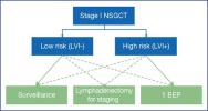
Conversely, the strategy can be adapted to the risk associated with the presence of LVI (Figure 1):
Figure 1.
Risk-appropriate strategy for the management of stage 1 NSGCT.
- In case of low-risk stage I NSGCT, the recurrence rate of 14% makes surveillance a primary option.
- In case of high-risk stage I NSGCT, the recurrence rate of 44% encourages the use of adjuvant chemotherapy with 1 BEP.
Whichever strategy is chosen, the disease-specific and overall survival rate is close to 99% [57,61].
The therapeutic strategy should be discussed in a MTM and the patient provided with information on all the therapeutic options and their risk-benefit ratio.
Table recommendation 3Recommendations for the management of stage 1 NSGCTLevels of recommendations for the management of stage 1 NSGCTStrength ratingRisk-appropriate treatment strategy (related to LVI) and surveillance without risk stratification are recommended options.StrongThe patient should be informed of the systematic and risk-appropriate approaches, along with the benefits and risks of the different options.StrongRisk-appropriate strategyLow risk (LVI-)Surveillance is the recommended option if the patient is compliantStrongIf the circumstances are not compatible with close surveillance adjuvant chemotherapy with 1 cycle of BEP is the recommended option.StrongHigh risk (LVI+)Adjuvant chemotherapy with 1 cycle of BEP is the recommended option given its proven superiority over staging lymphadenectomy. The patient should be informed of the benefits and risks of 1 cycle compared to 2.StrongIf ineligible for chemotherapy, surveillance is the recommended option.StrongLymphadenectomy for staging should be reserved for select cases in expert centres and nerve preservation should be practised.After lymphadenectomy, a pathological stage II should be discussed for surveillance or adjuvant chemotherapy.StrongAccording to the IGCCCG, in case of recurrence, and regardless of the initial treatment chosen, treatment consists of 3 or 4 cycles of BEP, followed by lymphadenectomy for residual masses if appropriateStrong
| Table recommendation 3 - Recommendations for the management of stage 1 NSGCT |
|
| Levels of recommendations for the management of stage 1 NSGCT
| Strength rating | | Risk-appropriate treatment strategy (related to LVI) and surveillance without risk stratification are recommended options. | Strong | | The patient should be informed of the systematic and risk-appropriate approaches, along with the benefits and risks of the different options. | Strong | | Risk-appropriate strategy | Low risk (LVI-) | Surveillance is the recommended option if the patient is compliant | Strong | | | | If the circumstances are not compatible with close surveillance adjuvant chemotherapy with 1 cycle of BEP is the recommended option. | Strong | | | High risk (LVI+) | Adjuvant chemotherapy with 1 cycle of BEP is the recommended option given its proven superiority over staging lymphadenectomy. The patient should be informed of the benefits and risks of 1 cycle compared to 2. | Strong | | | | If ineligible for chemotherapy, surveillance is the recommended option. | Strong | | | | Lymphadenectomy for staging should be reserved for select cases in expert centres and nerve preservation should be practised.
After lymphadenectomy, a pathological stage II should be discussed for surveillance or adjuvant chemotherapy. | Strong | | According to the IGCCCG, in case of recurrence, and regardless of the initial treatment chosen, treatment consists of 3 or 4 cycles of BEP, followed by lymphadenectomy for residual masses if appropriate | Strong |
|
|
| Treatment of testicular GCT at the metastatic stage |
The IS stage corresponds to patients with no lesions detectable on the TAP scan, whose marker levels do not decrease with half-life or increase after orchiectomy.
This context is suggestive of micro-metastatic disease if the contralateral testis is healthy and there is no differential diagnosis with non-specific elevation of the marker. The literature on systematic lymphadenectomy for staging reports a pathological stage II in 87% of these cases.
The treatment is the same as for metastatic GCT with a good prognosis.
|
|
Metastatic Seminoma (mSeminoma) |
|
|
Low volume mSeminoma (IIA - IIB < 3 cm) |
Diagnosis of a low-volume metastatic form of seminoma (especially stage IIA, ≤ 2 cm) is difficult, especially in the context of normal tumour markers. Treatment should only be initiated if there is diagnostic certainty, which may involve reassessment by CT scan at 6–8 weeks, or a biopsy.
Combination therapy studies are under way to limit morbidity in this context.
Radiotherapy is recommended for:
• | for the IIA stages: 30 Gy in dogleg technique
|
• | for the IIB stages: 30 Gy in dogleg technique including pathological adenopathies with a safety margin of 1–1.5 cm, and a boost of 6 Gy on the diseased area.
|
The relapse-free survival rate is 92 and 90% respectively [64].
Chemotherapy (3 BEP or 4 EP in case of contraindication to Bleomycin) is an alternative, with a different toxicity profile.
A quality meta-analysis suggested comparable efficacy with more acute toxicity after chemotherapy and late toxicity after radiotherapy (digestive, second cancers) [65]. Toxicity appeared to be more unfavourable with radiotherapy in high-volume IIB stages. No randomised studies have compared chemotherapy and radiotherapy in this indication.
|
|
Advanced mSeminoma (IIB ≥ 3 cm – IIC – III) |
There are limitations in the literature regarding metastatic seminoma [66].
Patients treated for advanced mSeminoma in the IGCCCG good prognosis group should receive chemotherapy with 3 BEP or 4 EP (if bleomycin is contraindicated). The disease-specific survival rate reported after 4 PE in this context is close to 100% [67].
Recent results from the SEMITEP cohort 2 de-escalation study (2 EP + 1 AUC7 carboplatin after an FDG PET-negative scan) in patients with a good prognosis stage IIB, IIC and III tumours, showed a 3-year relapse-free survival rate of 89.9% (79.6 – 95.3%) after a median follow-up of 33.9 months (95% CI: 30.2 – 39.3). Overall survival was close to 99% at 3 years. Relapse-free survival was similar to that indicated in the latest update of the IGCCCG International Classification of seminomas. The data should be confirmed before this regime becomes a standard for this population with a good prognosis [68].
Patients treated for advanced mSeminoma who are in the IGCCCG intermediate prognosis group should receive 4-BEP chemotherapy, by extrapolation from the practice regarding mNSGCT, although no randomised trial has targeted this population. The relapse-free survival rate reported after 4 VIP in this context is 83% [67].
Table recommendation 4Recommendations for the management of metastatic SeminomaStageRecommendationLevelLow volume metastatic seminomaPatients should be informed of the benefit-risk ratio of radiotherapy and chemotherapy in low-volume stage IIA and IIB.StrongWhen chemotherapy is chosen, 3 BEP or 4 EP provide the same level of oncological control.StrongAdvanced metastatic seminomaStarting from high-volume stage IIB, the choice of chemotherapy protocol is based on the IGCCCG prognostic group, according to the same modalities as NSGCT.Strong
| Table recommendation 4 - Recommendations for the management of metastatic Seminoma |
|
| Stage | Recommendation | Level | | Low volume metastatic seminoma | Patients should be informed of the benefit-risk ratio of radiotherapy and chemotherapy in low-volume stage IIA and IIB. | Strong | | | When chemotherapy is chosen, 3 BEP or 4 EP provide the same level of oncological control. | Strong | | Advanced metastatic seminoma | Starting from high-volume stage IIB, the choice of chemotherapy protocol is based on the IGCCCG prognostic group, according to the same modalities as NSGCT. | Strong |
|
|
|
Metastatic NSGCT (mNSGCT) |
|
|
mNSGCT in the IGCCCG good prognosis group |
The recommended treatment for mNSGCT in the IGCCCG good prognosis group is based on 3 cycles of BEP or 4 cycles of EP if bleomycin is contraindicated [69]. BEP is administered according to a 21-day cycle:
(B) Bleomycin30 mgD1 - D8 - D15(E) Etoposide100 mg/m2D1 to D5(P) Cisplatin20 mg/m2D1 to D5
|
| (B) Bleomycin | 30 mg | D1 - D8 - D15 | | (E) Etoposide | 100 mg/m2 | D1 to D5 | | (P) Cisplatin | 20 mg/m2 | D1 to D5 |
|
In special cases of mNSGCT in the IGCCCG stage IIA good prognosis group , lymphadenectomy or surveillance may be discussed if there is diagnostic doubt (absence of marker elevation).
In case of surveillance, biological examinations and imaging are repeated at 6 weeks. Target stability or growth, combined with markers that are still normal, orient toward teratoma and indicate lymphadenectomy [70]. The exceptional possibility of a pure embryonal carcinoma, whose rapid evolution requires primary chemotherapy, should not be overlooked.
In case of lymphadenectomy, adjuvant chemotherapy with 2 BEP [71] should be discussed if there are unfavourable histoprognostic criteria (number of nodes invaded, capsular rupture). The rate of complete remission is 98% [71].
In special cases of suspected recurrence of stage IIA with negative markers 2 years after the initial diagnosis, a radio-guided biopsy should be considered to confirm the diagnosis of germ cell tumour recurrence.
|
|
mNSGCT in the IGCCCG intermediate prognosis group |
The recommended treatment of mNSGCT in the IGCCCG intermediate prognosis group is based on 4 cycles of BEP.
In case of contraindication to bleomycin, patients are treated with 4 cycles of VIP every 21 days:
(V) Etoposide75 mg/m2D1 to D5(I) Ifosfamide1.2 g/m2D1 to D5(P) Cisplatin20 mg/m2D1 to D5Mesna1.5 g/m2D1 to D5
|
| (V) Etoposide | 75 mg/m2 | D1 to D5 | | (I) Ifosfamide | 1.2 g/m2 | D1 to D5 | | (P) Cisplatin | 20 mg/m2 | D1 to D5 | | Mesna | 1.5 g/m2 | D1 to D5 |
|
|
|
mNSGCT in the IGCCCG poor prognosis group |
Patients with mNSGCT with a poor prognosis according to IGCCCG should receive a first cycle of BEP. The decrease in markers according to their half-life should be evaluated between D18 and D21 of the 1st cycle. If the decrease is insufficient, survival is reduced [72]. A dedicated tool is available at: calculation-tumor/.
In the event of a favourable decrease, patients should receive 3 cycles of additional PET chemotherapy, 4 in total, possibly followed by surgery for residual masses.
In the event of an unfavourable decrease, patients should receive the intensified protocol according to the results of the GETUG 13 study [73]. This study showed significant improvement in progression-free survival (59% vs. 48%, HR 0.66 [0.44–1.00], p=0.05) between the intensified arm and 4 BEP, with a trend towards improvement in overall survival and most of all, a marked decrease in salvage treatment with intensive chemotherapy.
In case of a contraindication to bleomycin, patients are treated with 4 cycles of VIP.
Given the rarity and complexity of these oncological situations, it is recommended that patients be included in clinical trials and referred to expert centres. The volume of patients treated in a centre is correlated with the survival of patients with mNSGCT with an intermediate or poor prognosis [74].
Table recommendation 5Recommendations for the management of mNSGCTRecommendationStrength ratingIIA stages without marker elevation should suggest an unspecific picture, teratoma or embryonal carcinoma. Embryonal carcinoma can be excluded by biopsy or lymphadenectomy. In other cases, the biological and radiological re-evaluation at 6 weeks will assist with decision making.StrongmNSGCTs in the IGCCCG good prognosis group are eligible for chemotherapy with 3 BEP (or 4 EP).StrongmNSGCTs in the IGCCCG intermediate prognosis group are eligible for chemotherapy with 4 BEP (or 4 VIP).StrongmNSGCTs in the IGCCCG poor prognosis group are eligible for a first cycle of BEP. The ensuing approach is adapted to the reduction kinetics of the markers between D1 and D21.Strong
| Table recommendation 5 - Recommendations for the management of mNSGCT |
|
| Recommendation | Strength rating | | IIA stages without marker elevation should suggest an unspecific picture, teratoma or embryonal carcinoma. Embryonal carcinoma can be excluded by biopsy or lymphadenectomy. In other cases, the biological and radiological re-evaluation at 6 weeks will assist with decision making. | Strong | | mNSGCTs in the IGCCCG good prognosis group are eligible for chemotherapy with 3 BEP (or 4 EP). | Strong | | mNSGCTs in the IGCCCG intermediate prognosis group are eligible for chemotherapy with 4 BEP (or 4 VIP). | Strong | | mNSGCTs in the IGCCCG poor prognosis group are eligible for a first cycle of BEP. The ensuing approach is adapted to the reduction kinetics of the markers between D1 and D21. | Strong |
|
|
|
Evaluation of metastatic GCT in systemic treatment |
Serum tumour markers should be evaluated on day one of the first cycle of chemotherapy. This value is used to define the final IGCCCG prognostic group.
If there is a time lapse between orchidectomy and chemotherapy, the CT scan should be updated so that any progression before chemotherapy is not misinterpreted in the event of insufficient regression at re-evaluation during chemotherapy.
Serum tumour markers should be evaluated:
• | Every week during chemotherapy when they are elevated,
|
• | Every cycle when normal.
|
Marker kinetics between D1 and D21 should be evaluated in patients in the IGCCCG poor prognosis group (section III.D.3.c).
In the event of an increase in markers during chemotherapy treatment, generally after the 3rd or 4th cycle (a rare situation and more frequent in patients with a tumour with a poor prognosis), patients should be referred to a specialised centre for subsequent care.
A CT TAP should be performed after 2 cycles of chemotherapy if there is a strong suspicion that surgery to remove residual mass might be called for at a later date (initial volume > 3 cm and/or presence of teratoma in the initial primary tumour). In the event of tumour growth, chemotherapy is still completed if the biological progression is favourable.
|
|
At the end of chemotherapy |
A biological and radiological reassessment by CT TAP should be performed 3 to 4 weeks after the end of chemotherapy. The changes in the targets on imaging should be interpreted according to the RECIST 1.1 criteria [75].
Salvage chemotherapy is generally recommended in case of disease progression (increase in markers and diffuse disease) following 1st line chemotherapy.
|
|
Management of post-chemotherapy residual masses |
Reduction in residual mSeminoma masses is usually delayed. A new biological and radiological evaluation by CT TAP after 3 months is useful to assess the reduction in the targets.
In case of a residual mass < 3 cm, a viable tumour is almost never noted. Careful surveillance is recommended, especially since the desmoplastic rearrangements increase the morbidity associated with lymphadenectomy of residual masses in this context [76].
In case of a mass ≥3 cm, 18FDG PET is recommended. It should be performed 6 weeks after chemotherapy to reduce false positives due to inflammation. Hypermetabolism of the targets, suggestive of a viable tumour, is the indication for surgery [77]. The characteristics of 18FDG PET for this indication were evaluated by meta-analysis at 78%, 86%, 58%, 84% and 0.9, for sensitivity, specificity, positive predictive value, negative predictive value, precision and AUC, respectively [78]. If fixation is questionable, 18FDG PET or CT imaging should be repeated to assess changes in lesion size or fixation intensity.
Any residual mNSGCT mass that measures more than 1 cm in the minor axis should be surgically resected 4 to 6 weeks after the end of chemotherapy. There is no reliable preoperative predictor of residual tumour.
Subcentimetric masses contain necrosis, teratoma or another viable tumour component in 74.7%, 21.4% and 3.9% of cases respectively [79]. The risk-benefit ratio is not in favour of immediate systematic treatment. However, the possibility of late recurrence should not be neglected in extended surveillance.
18FDG PET has no place in the evaluation of residual mNSGCT masses due to the false negatives in chemo- and radio-resistant teratoma [80].
Lymphadenectomy for residual masses is a regional surgery and cannot be limited to the excision of macroscopically visible masses. It should be considered when the disease is stabilised, and when markers increase thought should be given to chemotherapy instead. Preoperative, multidisciplinary planning is essential. Combined procedures (sacrifice of adjacent organ, vascular surgery) are necessary in 13.5% [81] to 27.2°% of the cases [82].
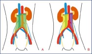
For radical lymphadenectomy the standard template is bilateral (Figure 2A). Dissection should be complete up to the anterior longitudinal ligament. The absence of lumbar vessel ligation and the persistence of a segment of spermatic cord are risk factors for recurrence. Nerves should be preserved whenever possible.
Figure 2.
Radical bilateral (A) and modified template (B) right unilateral (yellow) and left unilateral (purple) lymphadenectomy. The lower limit of lymphadenectomy is always the internal inguinal ring.
In order to reduce ejaculatory function loss in lymphadenectomy, a modified template has been proposed, aimed at preserving, at least unilaterally, the lumbar post-ganglionic sympathetic fibres running laterally to the aorta and the vena cava to converge around the origin of the inferior mesenteric artery and give rise to the superior hypogastric plexus.
The unilateral modified approach (regions shown in Figure 2B) should only be discussed for patients who strictly meet the following criteria: (i) mass initially and definitively ≤ 5 cm, (ii) ipsilateral to the primary tumour, (iii) without inter-aortic/caval mass [83].
Patients selected for modified lymphadenectomy should still be warned of the risk of retrograde ejaculation disorders and should be offered cryopreservation [76].
The laparoscopic approach which could be robot-assisted is the subject of publications by expert centres after evaluation in staging lymphadenectomy. Most of the series have a small population [84, 85], offer only limited follow-up [85], and often consist of a wide selection of cases (modified lymphadenectomy more frequent than radical lymphadenectomy, without associated procedures) [84]. Although the general postoperative advantages of the laparoscopic approach are undeniable, no compromise should be made in the completeness of the procedure, and surgery performed in non-expert centres may have led to unusual recurrences, possibly related to a non-optimal procedure (non-ligation of the lumbar vessels, for example) [86]. Given the possible difficulties of dissection with the need for complementary procedures (vascular, digestive-bone, urinary), these techniques should be reserved for centres with the expertise.
Table recommendation 6Recommendations for the management of mSGCTRecommendationStrength ratingIn case of a persistent residual mSGCT mass ≥ 3 cm, 18FDG PET performed 6 weeks after chemotherapy makes it possible to select patients for whom lymphadenectomy is suitable.IntermediateLymphadenectomy is suitable for any residual mSGCT mass greater than 1 cm in the smallest dimension. 18FDG PET has no place in patient selection.StrongLymphadenectomy for residual masses is envisaged when the markers are normalised or when their reduction kinetics is favourable.String
| Table recommendation 6 - Recommendations for the management of mSGCT |
|
| Recommendation | Strength rating | | In case of a persistent residual mSGCT mass ≥ 3 cm, 18FDG PET performed 6 weeks after chemotherapy makes it possible to select patients for whom lymphadenectomy is suitable. | Intermediate | | Lymphadenectomy is suitable for any residual mSGCT mass greater than 1 cm in the smallest dimension. 18FDG PET has no place in patient selection. | Strong | | Lymphadenectomy for residual masses is envisaged when the markers are normalised or when their reduction kinetics is favourable. | String |
|
|
|
Extra-peritoneal residual masses |
The discrepancy between the histology of retroperitoneal masses and that of masses at other metastatic sites is estimated to be 17% in case of retroperitoneal fibrosis but 42–58% in case of teratoma and 47–53% in case of viable germ cell tumours other than teratoma [87, 88]. No extrapolation is possible between the different sites. Surgical management should be discussed in multidisciplinary teams.
Unless there is a threatening lesion, the retroperitoneum is the first surgical site. The concomitant or sequential nature of interventions will depend on the expected morbidity [89].
In case of bilateral lung involvement, there is no need to explore the contralateral field if there is exclusive fibrosis or necrosis in the first field.
|
|
Histological result of residual mass lymphadenectomy |
Histological analysis of residual masses identifies:
• | for Seminoma, up to 30% tumour tissue (these are patients selected by 18FDG PET)
|
• | for NSGCT: teratoma in 40% of the cases, another active tumour component in 10% of the cases, and necrosis in 50% of the cases [ 76]. There is no systematic correlation between testicular and retroperitoneal histology: teratoma has been identified in 20% of the residual mass lymphadenectomies performed in patients treated by orchidectomy for pure embryonal carcinoma [ 90]. |
Only the presence of teratoma or necrosis in residual masses is not an indication for additional chemotherapy.
Surveillance, radiotherapy or adjuvant chemotherapy should be discussed in the presence of an active tumour. There is no standard recommendation. The risk factors for progression in this context are: incomplete resection, >10% viable tumour cells and an intermediate or poor IGCCCG prognosis [76].
|
| Post-chemotherapy tumour recurrences or chemotherapy-refractory disease |
Relapsed patients should be classified according to the International Classification of Relapsed Patients (Table 7), which is divided into 5 groups [91].
The management of tumour recurrence after chemotherapy or chemotherapy-refractory disease is based on salvage chemotherapy.
Several combinations of chemotherapy are recommended:
• | 4 cycles of VIP: Etoposide, ifosfamide, cisplatin
|
• | 4 cycles of TIP: Paclitaxel 250 mg/m2 on D1 continuously over 24 hours (non-MA), ifosfamide 1,500 mg/m2 from D2 to D5, cisplatin 25 mg/m2 from D2 to D5
|
• | 4 cycles of VeIP: Vinblastine 0,11 mg/kg on D1 and D2, Ifosfamide 1,200 mg/m2 from D1 to D5, Cisplatin 20 mg/m2 from D1 to D5
|
These protocols lead to a long-term remission rate of 15 to 50% [92] depending on prognostic factors that have been identified: location and histology of the primary tumour, response to the first-line treatment, duration of remission and the rate of markers on recurrence [91].
Intensive chemotherapy with peripheral stem cell support did not demonstrate superiority over salvage therapy in phase III trials, but an international retrospective analysis would appear to show improved survival in all prognostic subgroups [93].
The ongoing international TIGER study is evaluating standard salvage treatment with 4 cycles of TIP versus intensive chemotherapy according to the TICE protocol.
Gemcitabine has proven to be effective in the treatment of refractory disease by synergistic action with cisplatin [94].
It is strongly recommended that for salvage therapy, these rare patients should be referred to facilities that participate in clinical trials or that have experience in the management of relapses.
Late recurrence is defined as progressive relapse 2 years after curative treatment with chemotherapy initially effective on a metastatic disease [76]. It is infrequent and represents 1 to 3% of the cases. Massive adenopathies at the initial presentation and the presence of teratoma when the residual masses are removed are contributing factors [95]. The histology is divided into: viable germ cell tumour in 60–70% of the cases, teratoma in 60% of the cases or a malignant somatic transformation of the teratoma (sarcoma, adenocarcinoma) in 20% of the cases [76,95].
Late recurrences are a particular feature of GCT with a poorer prognosis and require optimal multidisciplinary management [95].
Surgery is the immediate treatment when the tumour markers are normal if the primary hypothesis is teratoma. In case of marker elevation, surgery is preceded by chemotherapy if the hypothesis is viable GCT, especially since recurrence is multifocal or difficult to resect [76,95]. Surgery should be complete, given the high probability of a chemoresistant component.
In case of a non-resectable lesion, biopsies should be taken to guide second-line systemic treatment. In case of seminoma, radiotherapy may be an alternative for limited non-resectable lesions [96].
The prognosis is negative, with one study reporting a progression-free survival at 3 years of 41% and an overall survival of 61% [97].
|
| Stage I germ cell tumour surveillance |
Monitoring of stage I GCT is based on clinical examination, serum assay of the 3 tumour markers and thoraco-abdomino-pelvic CT scan (Table 8).
Self-examination is recommended, given a relative risk of testicular cancer which is close to 25% in a population with a history of GCT. However, the rate of metachronous GCT is estimated at 1% [98].
The European Society of Urogenital Radiology (ESUR) recommends an annual ultrasound of the remaining testis if it has at least 5 microlithiases per field [10].
Only one randomised study has demonstrated the value of reducing the frequency of stage I NSGCT surveillance CT scans to 2 in the first year [99]. Otherwise, these are only expert opinions.
|
| Monitoring metastatic germ cell tumours in remission after chemotherapy |
The follow-up of patients with mGCT (Table 9) in remission after chemotherapy should address several concerns:
• | diagnosis of the relapse (mode, time, site, etc.)
|
• | long-term side effects
|
• | The toxicity of low doses of irradiation related to repeat examinations
|
|
|
Quality of life and long-term toxicity after testicular cancer treatment |
The immediate toxicity of chemotherapy (asthenia, nausea, vomiting, neurological toxicities, tinnitus) usually disappear after a few months.
The long-term toxicity of adjuvant BEP chemotherapy is poorly described but appears to be moderate or almost non-existent [100].
In the follow-up of these patients, it is important to reduce or eliminate risk factors (smoking, physical inactivity, overweight) early and to monitor blood pressure, lipid disorders and androgen function.
Metabolic syndrome may be a combination of high blood pressure, dyslipidaemia and endocrine disorders that over the long-term cause cardiovascular disease [101]. Cardiovascular disease-related mortality for patients who have been treated for a germ cell tumour is higher than in the general population [102].
Raynaud’s syndrome is related to cisplatin and/or bleomycin toxicity in the vessels and causes vascular spasms on exposure to cold.
The risk of deep vein thrombosis is increased in advanced mTGCT [103, 104]. There is no consensus on the predictive factors (LDH, AJCC stage, body surface area, size of retroperitoneal lymph node masses >35 mm). While thromboprophylaxis appears to be favourable in these contexts, studies with a high level of evidence are lacking. It is recommended to administer chemotherapy by peripheral veins when the venous network so allows.
In most instances, this is a sensory peripheral neuropathy resulting in paresthesias that occur in the extremities after cisplatin administration. Toxicity is observed in 29% of patients to varying degrees, often grade 1–2. It increases to 46% in patients who receive more than 5 cycles of cisplatin [105].
Ototoxicity is linked to cisplatin and most of the times results in tinnitus, more rarely in a decrease in hearing acuity, mainly for high frequencies of 4,000 Hz, and a hearing aid may be required in some cases.
The use of bleomycin may induce long-term toxicity which increases with smoking. Bleomycin is responsible for 1–3% of toxic deaths in GCT patients.
Cisplatin can induce long-term renal toxicity with a 20–30% loss of function. Impaired kidney function potentiates the toxicity of other products such as bleomycin.
Fatigue may persist for up to several months after the end of chemotherapy, and this is exacerbated by hypoandrogenism. Insufficient androgen production results in a decrease in bioavailable testosterone and an increase in LH secretion [105].
The risk of a second solid cancer in patients treated for GCT occurs more than 10 years after the end of treatment. The younger the patient, the more pronounced it is. The risk varies according to the type of treatment received between chemotherapy and radiotherapy and increases when both treatments are combined. It persists more than 35 years after the end of treatment. Radiotherapy induces tumours in the treated area (colon, stomach, pancreas, bladder, urinary tract) [106].
The risk of leukaemia is dependent on the administration of a dose of cisplatin and etoposide above 2 g/m2. In most instances, this is an acute myeloid or lymphoblastic leukaemia. The ratio between the monitored and the treated population is 2.6 (95% CI: 2.1 – 3.2), often discovered 10 years after treatment of GCT.
TGCT: testicular germ cell tumour
SGCT: seminomatous germ cell tumour
NSGCT: non-seminomatous germ cell tumour
mGCT: metastatic germ cell tumour
GCNIS: germ cell neoplasia in situ
CT: computed tomography
MRI: magnetic resonance imaging
AFP: alpha feto protein
LDH: lactate dehydrogenase
hCGt: total human chorionic gonadotropin
18FDG PET: positron emission tomography with 18-fluorodesoxyglucose
IGCCCG: international germ cell collaborative consensus group
Les tumeurs malignes du testicule sont dans 98 % des cas des tumeurs germinales (TG), réparties en TGS et TGNS dans 60 et 40 % des cas [1].
Les TG sont rares avec 2 769 nouveaux cas en France en 2018 contre 1 319 en 1990 [1]. Une augmentation de l’incidence est rapportée en France (+21,3 % entre 2000 et 2014) [2] et dans le monde, comme au Canada où elle est passée de 3,77/100 000 en 1971 à 7,06/100 000 en 2015 [3]. Une augmentation supplémentaire estimée de 28 % est attendue d’ici 2025 [4].
Le pic d’incidence se situe entre 30 et 34 ans [5], voire 35 ans [2], les séminomes se déclarant plus tardivement, vers 39 ans en moyenne.
Certains facteurs de risque de TG sont consensuels [6]: syndrome de dysgénésie testiculaire (cryptorchidie, hypospadias, hypo- ou infertilité), antécédent personnel ou familial (1er degré) de TG, atrophie testiculaire (< 12 ml). D’autres sont discutés comme la consommation intense de cannabis [7], les pesticides, les solvants organiques, la morphométrie. Une étude a rapporté un lien (odds ratio [OR] 3,32, p = 0,04) entre TG et antécédent familial maternel au premier degré de cancer ovarien [8] mais n’est pas concordante avec une étude de registre suédoise [9].
Les microlithiases testiculaires isolées ne doivent pas être considérées comme un facteur de risque, mais leur association à des facteurs de risque connus peut conduire à une surveillance échographique semestrielle ou annuelle et aboutir à une confrontation histologique en cas de lésion focale [10].
|
|
Diagnostic et évaluation préthérapeutique |
|
| Symptômes et contexte clinique |
Les TG sont le plus souvent diagnostiquées devant des symptômes locaux: palpation d’une masse scrotale, dure, asymptomatique, parfois au décours d’un épisode douloureux. Il n’y a pas de latéralité préférentielle. L’atteinte bilatérale synchrone est exceptionnelle. La maladie est localisée dans 60–70 % des cas.
Dans moins de 5 % des cas, le diagnostic sera évoqué devant des symptômes régionaux ou généraux: douleur ou masse abdominale, détresse respiratoire, gynécomastie, ganglion de Troisier [11].
|
|
Marqueurs tumoraux sériques |
Le dosage de trois marqueurs sériques est recommandé systématiquement avant et après orchidectomie, pour leur valeur respectivement diagnostique et pronostique. Des marqueurs négatifs n’éliminent pas le diagnostic. C’est leur valeur postopératoire qui est à la base des classifications pronostiques.
L’interprétation de leur cinétique nécessite la connaissance de leur demi-vie:
• | l’AFP est principalement produite par les tumeurs vitellines et le carcinome embryonnaire. Elle n’est pas sécrétée par le séminome ou le choriocarcinome. Sa demi-vie est de 5–7 jours. Elle n’est pas spécifique et peut être retrouvée en oncologie digestive (carcinome hépatocellulaire, pancréas, estomac) et dans certaines hépatopathies non tumorales;
|
• | la gonadotrophine chorionique humaine totale (hCGt) est systématiquement sécrétée par le choriocarcinome mais peut être retrouvée dans certains carcinomes embryonnaires et dans 10–20 % des séminomes. Sa demi-vie est de 24–36 heures. Son dosage peut apparaître élevé en cas d’élévation de la sous-unité ß de l’hormone lutéinisante (LH) ou de production pituitaire de ßhCG. Les classifications pronostiques reposent sur la valeur du dosage des hCG totales;
|
• | les LDH sont non spécifiques et corrélées au turn-over cellulaire et au volume tumoral.
|
La persistance de marqueurs élevés après orchidectomie sans lésion décelable au scanner (stade IS) peut indiquer la présence d’une maladie micrométastatique et nécessite une évaluation de la cinétique des marqueurs et une réévaluation scannographique avant décision d’une chimiothérapie.
Lorsqu’une chimiothérapie est indiquée pour métastase, seuls les marqueurs dosés en début de traitement oncologique permettent une classification précise selon l’IGCCCG, les marqueurs périopératoires étant sources de sous- et surstadification [12].
|
|
Autres examens biologiques |
Suivant le contexte, des dosages hormonaux (dont la testostéronémie, l’estradiolémie, les dosages sériques d’hormone folliculo-stimulante [FSH] et LH) peuvent être proposés. Des données rétrospectives suggèrent une baisse de la fonction endocrine du testicule résiduel après chirurgie et plus encore après chimiothérapie par BEP, pouvant nécessiter une supplémentation androgénique dans 11 à 15 % des cas [13].
De nouveaux biomarqueurs prometteurs sont en cours d’évaluation, comme les dosages sériques de micro-acide ribonucléique (ARN) (notamment miR-371a-3p [14]) lorsque le plateau technique s’y prête (Polymerase Chain Reaction [PCR]). Des résultats encourageants ont été publiés dans des séries rétrospectives pour discriminer les patients porteurs de tumeurs viables au sein de masses résiduelles [15] toutefois le manque de robustesse des données et les zones de chevauchement entre les groupes ne permettent pas de recommander l’usage de ce marqueur en alternative à la chirurgie.
L’écho-Doppler scrotal est recommandé systématiquement, permettant de distinguer les lésions intra- et extratesticulaires, et d’évoquer certaines lésions bénignes, avec une sensibilité > 95 % lorsqu’elle est couplée à l’examen clinique [16]. L’aspect échographique peut être évocateur de TGNS, volontiers plus hétérogène que les TGS (nodules hypoéchogènes plus ou moins confluents et vascularisés en Doppler couleur) [17].
Des nodules testiculaires non palpables ont été rapportés dans 2,9 % des échographies réalisées pour infertilité [18]. Dans cette population les tumeurs germinales sont possibles, mais plus rares, et il est important d’évoquer d’autres diagnostics, comme les tumeurs stromales à cellules de Leydig (7 % des lésions non palpables), vascularisées, le plus souvent bénignes quand elles sont de petite taille [19] et les tumeurs éteintes ou burned out [20]
L’imagerie par résonance magnétique (IRM) scrotale peut être utile dans les cas d’échographie équivoque, pour aider à déterminer la malignité avec une sensibilité, spécificité, précision respectivement de 100, 87,5, 92,8 % [21, 22].
L’échographie de contraste, l’élastographie ultrasonore sont en cours d’évaluation pour leur capacité diagnostique et ne sont pas recommandées en pratique courante [22, 23, 24].
|
|
Imagerie abdomino-pelvienne et thoracique |
Le scanner abdomino-pelvien est systématiquement recommandé pour l’évaluation du stade ganglionnaire rétro-péritonéal. Sa sensibilité est de 70–80 % pour une cible seuil définie à 8–10 mm. Il permet également la détection d’éventuelles métastases viscérales [20,25].
L’IRM abdominale peut être réalisée pour l’évaluation ganglionnaire en cas de contre-indication au scanner injecté ou de refus du patient de l’irradiation [26], avec un résultat équivalent au scanner.
Le scanner thoracique est l’examen le plus sensible pour la détection des métastases pulmonaires ou des adénopathies médiastinales. Sa spécificité est réduite par le taux de faux positifs (micronodules pulmonaires non spécifiques) [20]. En pratique, le scanner thoraco-abdomino-pelvien sera réalisé dans la grande majorité des cas.
|
|
Autres modalités d’imagerie |
L’imagerie cérébrale (tomodensitométrie [TDM] ou IRM) est recommandée en cas de symptômes neurologiques et chez les sujets à risque: choriocarcinome, mauvais pronostic selon l’IGCCCG (10 % de lésions cérébrales), métastases pulmonaires étendues, tumeur non séminomateuse primitive du médiastin antérieur. Seuls 1–2 % des patients présenteront des métastases cérébrales mais elles sont identifiées chez 40 % des patients décédant d’une maladie évolutive [27].
La TEP-18FDG n’est pas recommandée dans le bilan de stadification initiale des TG. Ses faux négatifs comprennent le tératome mature et les adénopathies infracentimétriques [17,20].
Les autres examens d’imagerie (scintigraphie osseuse, échographie hépatique…) seront proposés en fonction du contexte clinique.
Tableau de recommandation 1Recommandations de bilan paraclinique des TGExamensRecommandationsGradesMarqueurs tumoraux sériques (hCGt, AFP, LDH)SystématiqueFortÉchographie scrotaleSystématiqueFortScanner TAPSystématiqueFortIRM abdominaleEn cas de contre-indication au scanner, pour l’évaluation ganglionnaireFortImagerie cérébraleEn cas de symptômes ou chez le sujet à risqueFortScintigraphie osseuseEn cas de symptômesFortTEP-18FDGNon recommandéeFort
| Tableau de recommandation 1 - Recommandations de bilan paraclinique des TG |
|
| Examens | Recommandations | Grades | | Marqueurs tumoraux sériques (hCGt, AFP, LDH) | Systématique | Fort | | Échographie scrotale | Systématique | Fort | | Scanner TAP | Systématique | Fort | | IRM abdominale | En cas de contre-indication au scanner, pour l’évaluation ganglionnaire | Fort | | Imagerie cérébrale | En cas de symptômes ou chez le sujet à risque | Fort | | Scintigraphie osseuse | En cas de symptômes | Fort | | TEP-18FDG | Non recommandée | Fort |
|
|
| Diagnostic anatomo-pathologique |
|
|
Place des biopsies testiculaires |
La biopsie percutanée des TG du testicule est proscrite.
En cas de doute diagnostic, une orchidectomie partielle par voie inguinale pourra être discutée, notamment en cas de tumeur de petite taille et/ou non palpable. Une tumeur maligne est attendue dans 25 % des cas de lésion infracentimétrique chez les patients infertiles [28]. L’analyse extemporanée peut éviter l’orchidectomie totale pour tumeur bénigne [29].
Les biopsies du testicule controlatéral à la TG (réalisées par voie inguinale) ne sont pas systématiquement recommandées du fait de leur balance bénéfice-risque: la morbidité est de l’ordre de 3 %. Elles peuvent être discutées pour les patients à risque à savoir les sujets jeunes porteurs de microlithiases et présentant au moins un facteur de risque de TG (voir Chapitre I.B) [30].
|
|
Aspects chirurgicaux - orchidectomie |
L’orchidectomie élargie par voie inguinale est le traitement local de référence des TG du testicule. En cas de volume métastatique important et/ou symptomatique, la chimiothérapie sera le premier traitement. L’orchidectomie devra être réalisée ensuite, révélant la persistance de TG testiculaire viable dans 51,4 % des cas, composée dans 30,4 % des cas de tératome [31].
L’abord inguinal est impératif pour respecter le drainage lymphatique et permettre la ligature première du cordon à l’orifice inguinal interne. Une suture non résorbable permettra son identification en cas de curage ganglionnaire rétropéritonéal dont il est la limite inférieure [32].
Une prothèse testiculaire pourra être proposée dans le même temps ou à distance.
L’orchidectomie partielle par voie inguinale pour TG ne sera recommandée qu’en cas de testicule unique ou de tumeurs bilatérales synchrones. Ses indications sont restrictives: fonction endocrine normale, volume tumoral inférieur à 30 % du volume gonadique, diamètre de la tumeur inférieur à 2 cm. Des biopsies de pulpe en territoire sain seront pratiquées à la recherche de néoplasie germinale in situ (NGIS).
|
|
Analyse et compte-rendu histo-pathologique |
Le diagnostic définitif repose sur l’analyse de la pièce d’orchidectomie.
Les caractéristiques macroscopiques doivent y figurer: côté, taille du testicule, plus grand axe tumoral, aspect de l’épididyme, du cordon et de la vaginale, multifocalité. L’échantillonnage doit porter sur: toutes les zones macroscopiquement suspectes (un prélèvement par cm de lésion si elle est homogène; inclusion en totalité des tumeurs de moins de 2 cm), comportant également du parenchyme sain, le hile testiculaire, l’albuginée, l’épididyme, ainsi que des sections proximales et distales du cordon [33, 34].
L’analyse microscopique doit définir le type histologique selon la classification de l’Organisation mondiale de la santé (OMS) 2016 [35] et préciser l’infiltration lymphovasculaire; l’atteinte de l’albuginée, de la vaginale, du rete testis, de l’épididyme et du cordon spermatique; la présence de NGIS. L’analyse immunohistochimique est recommandée en cas de doute.
|
|
Classification Tumor, Node, Metastasis (TNM) et des marqueurs tumoraux sériques |
La classification TNM (Tableau 1) est établie à partir de l’analyse histologique et du bilan d’extension. Elle a été mise à jour en 2016 [36].
La classification des marqueurs (Tableau 2) repose sur le dosage postopératoire dans les formes localisées et à J1 du premier cycle dans les formes métastatiques.
|
|
Classification American Joint Committee on Cancer (AJCC) |
La classification AJCC (Tableau 3) [37] est une agrégation de la classification 2009 de la Tumor, Node, Metastasis Staging System (TNMS). Sa définition est indispensable à la prise en charge thérapeutique.
|
|
Classifications pronostiques |
Dans les stades localisés, les facteurs pronostiques sont utilisés pour définir la stratégie thérapeutique adjuvante adaptée au risque.
Dans les TGS, le grand axe tumoral > 4 cm et l’invasion du rete testis ont été considérés comme facteurs de risque de récidive [38]. Dans une analyse ultérieure, seule la taille tumorale a été confirmée significative: il n’existait pas de seuil mais un risque proportionnel à la taille de la tumeur, variant de 9 % pour 1 cm à 26 % pour 9 cm [39]. L’atteinte du rete testis n’a cependant pas pu être éliminée, des études prospectives ayant retenu les deux critères historiques [40].
Dans les TGNS, l’invasion lymphovasculaire (ILV) est le seul facteur prédictif de récidive (jusqu’à 50 %) en analyse multivariée. Le pourcentage élevé (> 50 %) de carcinome embryonnaire et l’index de prolifération peuvent être considérés comme facteurs de risque additionnels [41].
Dans les formes métastatiques, le groupe collaboratif international sur les tumeurs germinales (IGCCCG) a établi en 1997 une classification fondée sur les caractéristiques du site primitif de la tumeur, le taux des marqueurs et les sites métastatiques [42]. La classification définitive fait appel au dosage des marqueurs à J1 du premier cycle et définit le nombre de cycles de chimiothérapie (Tableau 4).
Une nouvelle classification internationale devrait être bientôt disponible tant pour les TGS que les TGNS. Les communications orales ont concerné: l’actualisation des résultats de survie sans progression et globale (Tableau 5 et Tableau 6) [43]; la prise en compte de l’atteinte pulmonaire et de l’âge dans le pronostic des TGNS; l’utilisation du seuil de 2,5N pour les LDH pour classer le pronostic de TGS en intermédiaire.
|
|
Prise en charge thérapeutique |
|
| Préservation de la fertilité |
Seuls 50 % des patients se présentant pour cryoconservation dans un contexte de TG ont une concentration de spermatozoïdes normale [44]. Différentes hypothèses étiologiques sont proposées parmi: syndrome de dysgénésie testiculaire, effets systémiques, endocriniens, immuns, troubles du développement testiculaire [45].
Le nombre de cycles de chimiothérapie et la réalisation d’un curage rétropéritonéal seraient des facteurs péjoratifs pour la fertilité postthérapeutique. Le taux d’éjaculation rétrograde rapporté étant respectivement de 1–9 %, 11–29 % et 75 % respectivement après curage de stadification, de masses résiduelles avec et sans préservation nerveuse [45]. Une étude prospective menée auprès des centres d’étude et de conservation des œufs et du sperme (CECOS) rapporte une altération de la numération et de la motilité spermatocytaire associée à un taux plus élevé d’aneuploïdie chez les patients traités par TG pendant 1 an après radiothérapie et 2 ans après chimiothérapie [46].
La cryoconservation de sperme au CECOS est recommandée, idéalement avant l’orchidectomie et impérativement avant toute chimiothérapie, radiothérapie, ou chirurgie rétro-péritonéale. Elle a une valeur médico-légale.
|
| Concertation pluridisciplinaire |
La présentation des dossiers de TG est recommandée de façon systématique après orchidectomie, et dans certains cas douteux avant chirurgie.
|
| Traitement des TG testiculaires de stade I |
En l’absence de traitement de la NGIS, le risque de nouvelle TG à 5 ans est de l’ordre de 50 % [47]. Une présentation claire du risque encouru à moyen terme permettra au patient de peser la balance entre le bénéfice d’un traitement complémentaire différé pour désir de paternité et le risque de s’exposer à une nouvelle lésion.
Le traitement de la NGIS isolée chez un patient possédant un testicule controlatéral fonctionnel et sain est l’orchidectomie [47].
Si l’orchidectomie ne peut être considérée (patient monorchide) par la réunion de concertation pluridisciplinaire (RCP), la radiothérapie scrotale par 16 à 20 Gy en fractions de 2 Gy permet de l’éradiquer [47]. Une dose minimale de 18 Gy a pu être recommandée [48]. Elle induit une infertilité et parfois un hypogonadisme [47].
Les TGS de stade IA/IB sont caractérisées par une normalisation des marqueurs tumoraux sériques (hCGt, AFP, LDH) après l’orchidectomie et par un scanner TAP ne mettant pas en évidence de lésion secondaire. Le scanner est parfois douteux pour des éléments ganglionnaires évoquant un stade II: la répétition de l’examen après 6–8 semaines aide alors au choix du traitement adjuvant.
Les études de surveillance systématique des séminomes de stade I objectivent un taux de récidive à 5 ans de 17,7 % [49]. Les récidives sont très majoritairement de bon pronostic, en situation sous-diaphragmatique. Jusqu’à 70 % des récidives sont de faible volume et peuvent être traitées par radiothérapie seule plutôt que par chimiothérapie adaptée au groupe pronostique IGCCCG [50].
Les facteurs de risque sont sujets à débat et portent une faible puissance dans la prédiction du pronostic de la TGS de stade I [51]. La surveillance semble donc de première intention, sous réserve d’une compliance parfaite du patient [52]. La survie spécifique et globale des TGS de stade I surveillés dans des centres experts est supérieure à 99 % [53].
|
|
Chimiothérapie adjuvante par carboplatine |
La chimiothérapie adjuvante repose sur un cycle unique de carboplatine AUC7 (pas d’autorisation de mise sur le marché [AMM] dans cette indication). Un cycle de carboplatine AUC7 n’est pas inférieur à une radiothérapie adjuvante (20 Gy, lombo-aortique), en termes de taux de récidive (5 % vs 4 %), de délai de récidive et de survie spécifique avec un recul médian de 5 ans [54].
Si la toxicité aiguë est très faible, les effets secondaires à long terme de cette chimiothérapie ne sont pas encore connus.
Le protocole recommandé est une irradiation lombo-aortique exclusive à la dose de 20 Gy. Il réduit le taux de récidive à 4 % [55].
Le taux d’effets secondaires à long terme (inférieur à 2 %) et le risque de second cancer doivent limiter les indications à des cas particuliers [56]. La radiothérapie adjuvante ne doit pas être proposée en première intention chez les jeunes patients.
Une attitude de surveillance de tous les patients traités pour une TGS de stade I serait licite devant un risque global de rechute ganglionnaire de 17,7 %.
Cependant ce risque peut augmenter jusqu’à 26 % selon la taille de la tumeur initiale [39]. Une stratégie adaptée au risque peut alors être adoptée, discutant un cycle adjuvant de carboplatine AUC7.
Quelle que soit la stratégie retenue, le taux de survie spécifique et globale est proche de 100 %.
La stratégie thérapeutique sera discutée en RCP et le patient devra recevoir l’information concernant l’ensemble des options thérapeutiques en présentant leur balance bénéfice-risque. Il s’agit d’une décision partagée avec le patient.
Tableau de recommandation 2Recommandations de prise en charge des TGS stade IGrades des recommandations de prise en charge des TGS stade IGradeLa surveillance est l’option recommandée en cas de compliance du patient.FortAucun traitement adjuvant n’est recommandé dans les formes de faible risque.FortLa chimiothérapie par un cycle de carboplatine AUC7 est l’option recommandée en cas d’indication de traitement adjuvant.FortLa radiothérapie lombo-aortique n’est pas recommandée en première intention en cas de traitement adjuvant.Fort
| Tableau de recommandation 2 - Recommandations de prise en charge des TGS stade I |
|
| Grades des recommandations de prise en charge des TGS stade I | Grade | | La surveillance est l’option recommandée en cas de compliance du patient. | Fort | | Aucun traitement adjuvant n’est recommandé dans les formes de faible risque. | Fort | | La chimiothérapie par un cycle de carboplatine AUC7 est l’option recommandée en cas d’indication de traitement adjuvant. | Fort | | La radiothérapie lombo-aortique n’est pas recommandée en première intention en cas de traitement adjuvant. | Fort |
|
La surveillance systématique des TGNS de stade I s’accompagne d’un taux de survie spécifique à 15 ans de 99,1 % [57].
Néanmoins elle expose à un risque de récidive global de 19 %, variant de 14 à 44 % selon l’absence ou la présence de l’ILV [58]. Ces rechutes nécessitent une chimiothérapie éventuellement associée à un curage de masses résiduelles. L’évolution survient dans 80 % des cas la première année, 12 % la seconde, 6 % la troisième, 1 % la quatrième et cinquième. Il s’agit d’une forme de bon pronostic IGCCCG dans 94 % des cas.
La chimiothérapie adjuvante par 2 cycles de BEP, introduite dans les années 1990, avait montré une réduction du taux de récidive à moins de 3 % (suivi moyen 7,9 ans) pour des TGNS de stade I de haut risque [59]. La toxicité à moyen terme est faible et la fertilité des patients peu altérée. Les toxicités à très long terme ne sont pas rapportées dans la littérature.
Un cycle unique de BEP a prouvé sa supériorité par rapport au curage de stadification en termes de réduction des récidives dans une cohorte de TGNS de stade I comprenant 43 % de haut risque (survie sans progression à 2 ans, 99,4 % vs 91,8 %; hazard ratio [HR] 7,9) [60]. Les données du Swedish and Norwegian Testicular Cancer Group (SWENOTECA) rapportent un taux de récidive des TGNS de stade I de haut risque après 1 BEP de 3,4 % après un suivi médian de 8,1 ans [61]. Les données récentes de Cullen et al. confirment la faisabilité d’un cycle de BEP dans une population à haut risque évolutif avec un taux de rechute de 3,1 % dont la moitié correspondait à un growing teratoma [62]. Les données à long terme manquent mais les effets secondaires dose-dépendant sont limités, ce qui augmente le rapport bénéfice-risque de la chimiothérapie adjuvante.
En cas de décision d’une chimiothérapie adjuvante, un cycle de BEP semble être la meilleure option.
|
|
Curage rétropéritonéal de stadification |
Vingt à 30 % des patients porteurs d’une TGNS stade I présentent en fait une atteinte rétropéritonéale pathologique (stade II) parmi lesquelles 30 % environ récidiveront sans chimiothérapie adjuvante. Dix pour cent des patients de stade I pathologique auront une évolution métastatique à distance [63].
Les critères de risque de récidive après curage, et donc les indications de chimiothérapie, sont aujourd’hui mal définis (emboles, carcinome embryonnaire dominant, effraction capsulaire étendue) et peu applicables en pratique clinique.
L’infériorité du curage exclusif face à un cycle unique de BEP appelle à la réduction des indications (tératome pur, contingent extra-gonadique associé). En cas de réalisation de celui-ci, la limitation de la morbidité implique l’expertise du curage rétropéritonéal, des limites du territoire de curage modifié, de la préservation des branches nerveuses sympathiques donnant naissance au plexus hypogastrique supérieur.
La surveillance systématique des TGNS de stade I peut être appliquée sous réserve de l’information complète du patient vis-à-vis du risque de récidive (jusqu’à 44 % dans les formes de haut risque) motivant des traitements complémentaires de rattrapage plus morbides que le traitement adjuvant [57].
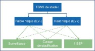
À l’inverse, la stratégie peut être adaptée au risque lié à la présence de l’ILV (Figure 1):
Figure 1.
Stratégie de prise en charge des TGNS de stade I adaptée au risque
• | en cas de TGNS de stade I de faible risque, le taux de récidive de 14 % fait de la surveillance une option de choix;
|
• | en cas de TGNS de stade I de haut risque, le taux de récidive de 44 % fait privilégier la chimiothérapie adjuvante par 1 BEP.
|
Quelle que soit la stratégie retenue, le taux de survie spécifique et globale est proche de 99 % [57,61].
La stratégie thérapeutique sera discutée en RCP et le patient devra recevoir l’information concernant l’ensemble des options thérapeutiques en présentant leur balance bénéfice-risque.
|
| Traitement des TG testiculaires de stade métastatique |
Le stade IS correspond aux patients sans lésion décelable au scanner TAP, dont le taux de marqueurs ne diminue pas selon la demi-vie ou augmente après l’orchidectomie.
Cette situation est évocatrice de maladie micro-métastatique, si le testicule controlatéral est sain et qu’il n’existe pas de diagnostic différentiel à l’élévation non spécifique du marqueur. La littérature des curages systématiques de stadification rapporte un stade II pathologique dans 87 % de ces cas.
Le traitement est celui d’une TG métastatique de bon pronostic.
|
|
TGSm de faible volume (IIA - IIB < 3 cm) |
Le diagnostic d’une forme métastatique de faible volume de séminome (notamment stade IIA, ≤ 2 cm) est difficile, surtout dans un contexte de marqueurs tumoraux normaux. Le traitement ne doit être initié qu’en cas de certitude diagnostique, ce qui peut impliquer une réévaluation par scanner à 6–8 semaines ou une biopsie.
Des études de combinaisons thérapeutiques sont en cours pour limiter la morbidité dans ce contexte.
La radiothérapie est recommandée pour:
• | les stades IIA: 30 Gy en crosse de hockey;
|
• | les stades IIB: 30 Gy en crosse de hockey intégrant les adénopathies pathologiques avec une marge de sécurité de 1–1,5 cm, et un boost de 6 Gy sur la zone pathologique.
|
Le taux de survie sans récidive est respectivement de 92 et 90 % [64].
La chimiothérapie (3 BEP ou 4 EP en cas de contreindication à la bléomycine) est une alternative, avec un profil de toxicité différente.
Une méta-analyse de qualité évoquait une efficacité comparable avec davantage de toxicités aiguës après chimiothérapie et tardives après radiothérapie (digestive, seconds cancers) [65]. Les toxicités semblaient davantage défavorables à la radiothérapie dans les stades IIB de haut volume. Aucune étude randomisée n’a comparé chimiothérapie et radiothérapie dans cette indication.
|
|
TGSm avancées (IIB ≥ 3 cm – IIC – III) |
Les données de la littérature concernant le séminome métastatique ont des limites [66].
Tableau de recommandation 3Recommandations de prise en charge des TNGS de stade IGrades des recommandations de prise en charge des TGNS de stade IGradeLa stratégie de traitement adaptée au risque (lié à l’ILV) ou la surveillance sans stratification du risque sont des options recommandées.FortLe patient doit recevoir l’information concernant les attitudes systématiques ou adaptées au risque, avec une présentation des bénéfices et risques des différentes options.FortStratégie adaptée au risqueFaible risque (ILV-)La surveillance est l’option recommandée en cas de compliance du patient.FortSi les conditions de surveillance rapprochée ne sont pas réunies, la chimiothérapie adjuvante par un cycle de BEP est l’option recommandée.FortHaut risque (ILV+)La chimiothérapie adjuvante par un cycle de BEP est l’option recommandée étant donné sa supériorité prouvée sur le curage de stadification. Le patient doit recevoir une information concernant les avantages et risques d’un cycle par rapport à deux.FortEn cas d’inéligibilité à la chimiothérapie, la surveillance est l’option recommandée.FortLe curage de stadification doit être réservé à des cas sélectionnés, dans des centres experts avec préservation nerveuse.Après curage, un stade II pathologique doit faire discuter une surveillance ou une chimiothérapie adjuvante.FortEn cas de récidive, et quelle que soit l’option thérapeutique initiale, le traitement consistera en trois ou quatre cycles de BEP selon la classification IGCCCG, suivis d’un éventuel curage de masses résiduellesFort
| Tableau de recommandation 3 - Recommandations de prise en charge des TNGS de stade I |
|
| Grades des recommandations de prise en charge des TGNS de stade I
| Grade | | La stratégie de traitement adaptée au risque (lié à l’ILV) ou la surveillance sans stratification du risque sont des options recommandées. | Fort | | Le patient doit recevoir l’information concernant les attitudes systématiques ou adaptées au risque, avec une présentation des bénéfices et risques des différentes options. | Fort | | Stratégie adaptée au risque | Faible risque (ILV-) | La surveillance est l’option recommandée en cas de compliance du patient. | Fort | | | | Si les conditions de surveillance rapprochée ne sont pas réunies, la chimiothérapie adjuvante par un cycle de BEP est l’option recommandée. | Fort | | | Haut risque (ILV+) | La chimiothérapie adjuvante par un cycle de BEP est l’option recommandée étant donné sa supériorité prouvée sur le curage de stadification. Le patient doit recevoir une information concernant les avantages et risques d’un cycle par rapport à deux. | Fort | | | | En cas d’inéligibilité à la chimiothérapie, la surveillance est l’option recommandée. | Fort | | | | Le curage de stadification doit être réservé à des cas sélectionnés, dans des centres experts avec préservation nerveuse.
Après curage, un stade II pathologique doit faire discuter une surveillance ou une chimiothérapie adjuvante. | Fort | | En cas de récidive, et quelle que soit l’option thérapeutique initiale, le traitement consistera en trois ou quatre cycles de BEP selon la classification IGCCCG, suivis d’un éventuel curage de masses résiduelles | Fort |
|
Les patients traités pour TGSm avancée du groupe bon pronostic IGCCCG doivent recevoir une chimiothérapie par 3 BEP ou 4 EP (en cas de contre-indication à la bléomycine). Le taux de survie spécifique rapporté après 4 EP dans cette situation est proche de 100 % [67].
Les résultats récents de l’étude de désescalade SEMITEP cohorte 2 (2 EP + 1 carboplatine AUC7 après négativation du PET scanner au FDG) chez les patients ayant une tumeur de bon pronostic de stade IIB, IIC et III, montrent une survie sans rechute à 3 ans de 89,9 % (79,6–95,3%) après une médiane de suivi de 33,9 mois (95 % intervalle de confiance [IC]: 30,2–39,3). La survie globale est proche de 99 % à 3 ans. La survie sans rechute est similaire à la dernière mise à jour de la classification internationale de l’IGCCCG des séminomes. Les données sont à confirmer avant que ce schéma ne devienne un standard pour cette population de bon pronostic [68].
Les patients traités pour TGSm avancée du groupe pronostic intermédiaire IGCCCG doivent recevoir une chimiothérapie par 4 BEP, par extrapolation de la pratique des TGNSm, alors qu’aucun essai randomisé n’a ciblé cette population. Le taux de survie sans récidive rapporté après 4 VIP dans cette situation est de 83 % [67].
Tableau de recommandation 4Recommandations de prise en charge des TGSmStadesRecommandationsGradesTGSm de faible volumeLes patients doivent recevoir une information concernant la balance bénéfice-risque de la radiothérapie et de la chimiothérapie dans les stades IIA et IIB de faible volume.FortLorsque la chimiothérapie est retenue, 3 BEP ou 4 EP procurent le même niveau de contrôle carcinologique.FortTGSm avancéeÀ partir des stades IIB de haut volume, le choix du protocole de chimiothérapie repose sur le groupe pronostique IGCCCG, selon les mêmes modalités que les TGNS.Fort
| Tableau de recommandation 4 - Recommandations de prise en charge des TGSm |
|
| Stades | Recommandations | Grades | | TGSm de faible volume | Les patients doivent recevoir une information concernant la balance bénéfice-risque de la radiothérapie et de la chimiothérapie dans les stades IIA et IIB de faible volume. | Fort | | | Lorsque la chimiothérapie est retenue, 3 BEP ou 4 EP procurent le même niveau de contrôle carcinologique. | Fort | | TGSm avancée | À partir des stades IIB de haut volume, le choix du protocole de chimiothérapie repose sur le groupe pronostique IGCCCG, selon les mêmes modalités que les TGNS. | Fort |
|
|
|
TGNS métastatiques (TGNSm) |
|
|
TGNSm du groupe IGCCCG bon pronostic |
Le traitement recommandé des TGNSm du groupe bon pronostic IGCCCG repose sur 3 cycles de BEP ou 4 cycles d’EP en cas de contre-indication à la bléomycine [69]. Le mode d’administration du BEP repose sur un cycle de 21 jours:
(B) Bléomycine30 mgJ1 - J8 - J15(E) Étoposide100 mg/m2J1 à J5(P) Cisplatine20 mg/m2J1 à J5
|
| (B) Bléomycine | 30 mg | J1 - J8 - J15 | | (E) Étoposide | 100 mg/m2 | J1 à J5 | | (P) Cisplatine | 20 mg/m2 | J1 à J5 |
|
Dans le cas particulier des TGNSm du groupe bon pronostic IGCCCG de stade IIA , un curage ganglionnaire ou une surveillance peuvent être discutés s’il existe un doute diagnostic (absence d’élévation des marqueurs).
En cas de surveillance, la biologie et l’imagerie seront répétées à 6 semaines. La stabilité ou la croissance des cibles, associée à des marqueurs toujours normaux orientera vers le tératome et indiquera un curage [70]. Il ne faudra pas méconnaître l’exceptionnelle possibilité d’un carcinome embryonnaire pur dont l’évolution rapide nécessite une chimiothérapie première.
En cas de curage ganglionnaire, une chimiothérapie adjuvante par 2 BEP [71] doit être discutée en présence de critères histopronostiques défavorables (nombre de ganglions envahis, rupture capsulaire). Le taux de rémission complète est de 98 % [71].
Dans le cas particulier des suspicions de récidive de stade IIA à marqueurs négatifs après 2 ans du diagnostic initial , une biopsie radioguidée doit être considérée pour confirmer le diagnostic de rechute de tumeur germinale.
|
|
TGNSm du groupe IGCCCG pronostic intermédiaire |
Le traitement recommandé des TGNSm du groupe IGCCCG pronostic intermédiaire repose sur 4 cycles de BEP.
En cas de contre-indication à la bléomycine, les patients seront traités par 4 cycles de VIP tous les 21 jours:
(V) Étoposide75 mg/m2J1 à J5(I) Ifosfamide1,2 g/m2J1 à J5(P) Cisplatine20 mg/m2J1 à J5Mesna1,5 g/m2J1 à J5
|
| (V) Étoposide | 75 mg/m2 | J1 à J5 | | (I) Ifosfamide | 1,2 g/m2 | J1 à J5 | | (P) Cisplatine | 20 mg/m2 | J1 à J5 | | Mesna | 1,5 g/m2 | J1 à J5 |
|
|
|
TGNSm du groupe IGCCCG mauvais pronostic |
Les patients atteints d’une TGNSm de mauvais pronostic selon l’IGCCCG doivent recevoir un premier cycle de BEP. La décroissance des marqueurs selon leur demi-vie doit être évaluée entre le J18 et le J21 du 1er cycle. En cas de décroissance insuffisante, la survie est diminuée [72]. Un outil dédié est disponible: calculation-tumor/.
En cas de décroissance favorable, les patients doivent recevoir 3 cycles de chimiothérapie supplémentaire par BEP, soit 4 au total, suivis d’une éventuelle chirurgie des masses résiduelles.
En cas de décroissance défavorable, les patients doivent recevoir le protocole intensifié selon les résultats du Groupe d’étude des tumeurs urogénitales (GETUG) 13 [73]. Cette étude a montré une amélioration significative de la survie sans progression (59 % vs 48 %, HR 0,66 [0,44–1,00], p = 0,05) entre le bras intensifié et 4 BEP, avec une tendance à l’amélioration de la survie globale et surtout à une nette diminution du traitement de rattrapage par chimiothérapie intensive.
En cas de contre-indication à la bléomycine, les patients recevront un traitement par 4 cycles de VIP.
Face à la rareté et la complexité de ces situations oncologiques, il est recommandé d’inclure les patients dans des essais cliniques et de les orienter vers des centres expérimentés. Le volume de malades traités dans un centre est corrélé à la survie des malades atteints d’une TGNSm de pronostic intermédiaire ou mauvais [74].
Tableau de recommandation 5Recommandations de prise en charge des TGNSmRecommandationsGradesLes stades IIA sans élévation des marqueurs doivent faire évoquer une image aspécifique, un tératome ou un carcinome embryonnaire. Ce dernier peut être exclu par biopsie ou curage. Dans les autres cas, la réévaluation biologique et radiologique à 6 semaines aidera la prise de décision.FortLes TGNSm du groupe IGCCCG de bon pronostic relèvent d’une chimiothérapie par 3 BEP (ou 4 EP).FortLes TGNSm du groupe IGCCCG de pronostic intermédiaire relèvent d’une chimiothérapie par 4 BEP (ou 4 VIP).FortLes TGNSm du groupe IGCCCG de mauvais pronostic relèvent d’un premier cycle de BEP. L’attitude ultérieure est adaptée à la cinétique de décroissance des marqueurs entre J1 et J21.Fort
| Tableau de recommandation 5 - Recommandations de prise en charge des TGNSm |
|
| Recommandations | Grades | | Les stades IIA sans élévation des marqueurs doivent faire évoquer une image aspécifique, un tératome ou un carcinome embryonnaire. Ce dernier peut être exclu par biopsie ou curage. Dans les autres cas, la réévaluation biologique et radiologique à 6 semaines aidera la prise de décision. | Fort | | Les TGNSm du groupe IGCCCG de bon pronostic relèvent d’une chimiothérapie par 3 BEP (ou 4 EP). | Fort | | Les TGNSm du groupe IGCCCG de pronostic intermédiaire relèvent d’une chimiothérapie par 4 BEP (ou 4 VIP). | Fort | | Les TGNSm du groupe IGCCCG de mauvais pronostic relèvent d’un premier cycle de BEP. L’attitude ultérieure est adaptée à la cinétique de décroissance des marqueurs entre J1 et J21. | Fort |
|
|
|
Évaluation des TG métastatiques en traitement systémique |
Les marqueurs tumoraux sériques doivent être évalués au premier jour du premier cycle de chimiothérapie. Cette valeur sera retenue pour définir le groupe pronostic IGCCCG définitif.
En cas de délai entre l’orchidectomie et la chimiothérapie, le scanner doit être actualisé pour qu’une progression avant chimiothérapie ne soit pas mal interprétée en cas de régression insuffisante à la réévaluation en cours de chimiothérapie.
|
|
En cours de chimiothérapie |
Les marqueurs tumoraux sériques doivent être évalués:
• | toutes les semaines en cours de chimiothérapie lorsqu’ils sont élevés;
|
• | à chaque cycle en cas de normalité.
|
La cinétique des marqueurs entre J1 et J21 doit être évaluée chez les patients du groupe mauvais pronostic IGCCCG (voir Chapitre III.D.3.c).
En cas d’augmentation des marqueurs sous chimiothérapie, en général après le 3e ou 4e cycle (situation rare et plus fréquente chez les patients ayant une tumeur de mauvais pronostic), les patients doivent être orientés vers un centre spécialisé pour la suite de la prise en charge.
Le scanner TAP doit être réalisé après 2 cycles de chimiothérapie en cas de forte suspicion d’une chirurgie de masses résiduelles à venir (volume initial > 3 cm et/ou présence de tératome dans la tumeur primitive initiale). En cas de croissance tumorale, la chimiothérapie sera tout de même conduite à son terme si l’évolution biologique est favorable.
Une réévaluation biologique et radiologique par scanner TAP doit être réalisée 3 à 4 semaines après la fin de la chimiothérapie. L’évolution des cibles à l’imagerie doit être interprétée selon les critères Response Evaluation Criteria in Solid Tumours (RECIST) 1.1 [75].
Une chimiothérapie de rattrapage est le plus souvent recommandée en cas de progression de la maladie (augmentation des marqueurs et maladie diffuse) à l’issue de la chimiothérapie de 1re ligne.
|
|
Prise en charge des masses résiduelles postchimiothérapie |
|
|
Masses résiduelles des TGS |
La régression des masses résiduelles de TGSm est habituellement retardée. Une nouvelle évaluation biologique et radiologique par scanner TAP après 3 mois est utile pour juger la régression des cibles.
En cas de masse résiduelle < 3 cm, il n’est quasiment jamais observé de tumeur viable. Une surveillance attentive est recommandée, ce d’autant que les remaniements desmoplastiques rendent davantage morbide le curage des masses résiduelles dans cette situation [76].
En cas de masse ≥ 3 cm, la TEP-18FDG est recommandée. Elle doit être réalisée 6 semaines après la chimiothérapie pour réduire les faux positifs inflammatoires. L’hypermétabolisme des cibles, évocateur de tumeur viable, pose l’indication chirurgicale [77]. Les caractéristiques de la TEP-18FDG dans cette indication ont été évaluées par méta-analyse à respectivement 78, 86, 58, 84 et 0,9 % pour la sensibilité, la spécificité, la valeur prédictive positive, la valeur prédictive négative, la précision et l’AUC [78]. En cas de fixation douteuse il est recommandé de refaire une imagerie par TEP-18FDG ou scanner pour évaluer l’évolution de la taille des lésions ou de l’intensité de fixation.
|
|
Masses résiduelles de TGNS |
Toute masse résiduelle de TGNSm mesurant plus d’un centimètre de petit axe doit faire l’objet d’une résection chirurgicale 4 à 6 semaines après la fin de la chimiothérapie. Il n’existe aucun élément préopératoire fiable de prédiction de la présence de tumeur résiduelle.
Les masses infracentimétriques contiennent de la nécrose, du tératome, un autre contingent tumoral viable dans respectivement 74,7, 21,4 et 3,9 % des cas [79]. La balance bénéfice-risque n’est pas en faveur d’un traitement systématique immédiat. La surveillance prolongée ne doit par contre pas négliger la possibilité d’une récidive tardive.
La TEP-18FDG n’a pas sa place dans l’évaluation des masses résiduelles de TGNSm du fait des faux négatifs du tératome chimio- et radioresistant [80].
Le curage de masses résiduelles est une chirurgie régionale et ne peut se limiter à l’exérèse des masses macroscopiquement visibles. Elle doit s’envisager lorsque la maladie est stabilisée et des marqueurs en augmentation doivent plutôt faire considérer une chimiothérapie. La planification préopératoire, multidisciplinaire, est essentielle. Les gestes combinés (sacrifices d’organe de voisinage, chirurgie vasculaire) sont nécessaires dans 13,5 % [81] à 27,2 % des cas [82].
Le territoire standard est bilatéral (Figure 2A) pour un curage radical. La dissection doit être complète jusqu’au ligament vertébral antérieur. L’absence de ligature des vaisseaux lombaires et la persistance d’un segment de cordon spermatique sont des facteurs de risque de récidive. Une préservation nerveuse doit être réalisée lorsqu’elle est possible.
Figure 2.
Territoire du curage bilatéral complet (A) et modifié (B) unilatéral droit (jaune) et gauche (violet). La limite inférieure du curage est toujours l’orifice inguinal interne.
Afin de réduire la morbidité éjaculatoire du curage, un territoire modifié a été proposé, visant à préserver au moins unilatéralement les fibres sympathiques postganglionnaires lombaires cheminant latéralement à l’aorte et la veine cave pour converger autour de l’origine de l’artère mésentérique inférieure et donner naissance au plexus hypogastrique supérieur.
L’approche modifiée unilatérale (territoires représentés sur la Figure 2B) ne doit se discuter que chez un patient répondant strictement aux critères suivants: (i) masse initialement et définitivement ≤ 5 cm, (ii) homolatérale à la tumeur primitive, (iii) sans masse inter-aortico-cave [83].
Les patients sélectionnés pour un curage modifiés doivent tout de même être avertis du risque de troubles de l’éjaculation rétrograde et une cryopréservation doit leur être proposée [76].
La voie d’abord laparoscopique éventuellement robot-assistée fait l’objet de publications par des centres experts après évaluation en situation de curage de stadification. Les séries sont majoritairement d’effectif faible [84, 85], n’offrent qu’un recul limité [85] et reflètent souvent une grande sélection des cas (curage modifié plus fréquent que radical, sans geste associé) [84]. Si les avantages généraux postopératoires de la voie d’abord laparoscopique sont indéniables, il ne faut accepter aucun compromis dans l’exhaustivité du geste, et des chirurgies pratiquées dans des centres non experts ont pu conduire à des récidives inhabituelles, possiblement en lien avec un geste non optimal (non-ligature des lombaires par exemple) [86]. Étant donné les difficultés possibles de dissection avec nécessité de gestes complémentaires (vasculaire, digestif osseux, urinaire), ces techniques doivent être réservées aux centres en ayant l’expertise.
Tableau de recommandation 6Recommandations de prise en charge des TGSmRecommandationsGradesEn cas de masse résiduelle persistante de TGSm ≥ 3 cm, la TEP-18FDG, réalisée 6 semaines après chimiothérapie, permettra de sélectionner les patients relevant d’un curage.ModéréToute masse résiduelle de TGNSm supérieure à 1 cm de petit axe relève d’un curage. La TEP-18FDG n’a pas de place dans la sélection des patients.FortLe curage de masses résiduelles s’envisage alors que les marqueurs sont normalisés ou que leur cinétique de décroissance est favorable.Fort
| Tableau de recommandation 6 - Recommandations de prise en charge des TGSm |
|
| Recommandations | Grades | | En cas de masse résiduelle persistante de TGSm ≥ 3 cm, la TEP-18FDG, réalisée 6 semaines après chimiothérapie, permettra de sélectionner les patients relevant d’un curage. | Modéré | | Toute masse résiduelle de TGNSm supérieure à 1 cm de petit axe relève d’un curage. La TEP-18FDG n’a pas de place dans la sélection des patients. | Fort | | Le curage de masses résiduelles s’envisage alors que les marqueurs sont normalisés ou que leur cinétique de décroissance est favorable. | Fort |
|
|
|
Masses résiduelles extra-péritonéales |
La discordance entre l’histologie des masses rétropéritonéales et celle des masses des autres sites métastatiques est estimée à 17 % en cas de fibrose dans le rétropéritoine mais 42–58 % en cas de tératome et 47–53 % en cas de tumeur germinale viable autre que le tératome [87, 88]. Aucune extrapolation n’est possible entre les différents sites. La prise en charge chirurgicale doit être discutée au sein d’équipes pluridisciplinaires.
Sauf lésion menaçante, le rétropéritoine est le premier site à opérer. Le caractère concomitant ou séquentiel des interventions sera fonction de la morbidité attendue [89].
En cas d’atteinte pulmonaire bilatérale, il n’y a pas de nécessité d’explorer le champ controlatéral en cas de fibrose ou nécrose exclusive dans le premier champ.
|
|
Résultat histologique du curage des masses résiduelles |
L’analyse histologique des masses résiduelles identifie:
• | pour les TGS, jusqu’à 30 % de tissu tumoral (il s’agit de patients sélectionnés par TEP-18FDG);
|
• | pour les TGNS: du tératome dans 40 % des cas, un autre contingent tumoral actif dans 10 % des cas, et de la nécrose dans 50 % des cas [ 76]. La corrélation n’est pas systématique entre histologie testiculaire et rétropéritonéale: il a pu être identifié du tératome dans 20 % des curages de masse résiduelle réalisé chez des patients traités par orchidectomie pour carcinome embryonnaire pur [ 90]. |
La présence exclusive de tératome ou de nécrose au sein des masses résiduelles n’indique pas de chimiothérapie complémentaire.
La présence de tumeur active doit faire discuter surveillance, radiothérapie ou chimiothérapie adjuvante. Il n’existe pas de standard de recommandation. Les facteurs de risque de progression dans ce contexte sont: une résection incomplète, > 10 % de cellules tumorales viables, un pronostic IGCCCG intermédiaire ou mauvais [76].
|
| Récidives tumorales postchimiothérapie ou maladie réfractaire à la chimiothérapie |
Les patients en récidive doivent être classés selon la classification internationale des patients en rechute (Tableau 7), répartis en 5 groupes [91].
La prise en charge d’une récidive tumorale après chimiothérapie ou d’une maladie réfractaire à la chimiothérapie repose sur la chimiothérapie de rattrapage.
Plusieurs combinaisons de chimiothérapie sont recommandées:
• | 4 cycles de VIP: Étoposide, ifosfamide, cisplatine;
|
• | 4 cycles de TIP: Paclitaxel 250 mg/m2 à J1 en continu sur 24 heures (hors AMM), ifosfamide 1 500 mg/m2 J2 à J5, cisplatine 25 mg/m2 J2 à J5;
|
• | 4 cycles de VeIP: Vinblastine 0,11 mg/kg J1 et J2, ifosfamide 1 200 mg/m2 J1 à J5, cisplatine 20 mg/m2 J1 à J5.
|
Ces protocoles conduisent à un taux de rémission à long terme de 15 à 50 % [92] selon des facteurs pronostiques identifiés: localisation et histologie de la tumeur primitive, réponse à la première ligne de traitement, durée de la rémission, taux des marqueurs à la récidive [91].
La chimiothérapie intensive avec support de cellules souches périphériques n’a pas prouvé de supériorité par rapport à un traitement de rattrapage dans les études de phase III mais une analyse rétrospective internationale semblerait montrer une amélioration de la survie dans tous les sous-groupes pronostiques [93].
L’étude internationale TIGER, en cours, évalue le traitement standard de rattrapage par 4 cycles de TIP versus une chimiothérapie intensive selon le protocole taxol, ifosfamide, carboplatine et étoposide (TICE).
La gemcitabine a prouvé une efficacité dans le traitement de maladie réfractaire par une action synergique avec le cisplatine [94].
Il est fortement recommandé en situation de rattrapage d’adresser ces rares malades dans les structures incluant dans les essais cliniques ou ayant l’expérience de la prise en charge des rechutes.
La récidive tardive est définie comme une reprise évolutive 2 ans après un traitement curatif par chimiothérapie initialement efficace sur une maladie métastatique [76]. Elle est peu fréquente, représentant 1 à 3 % des cas et favorisée par des adénopathies massives à la présentation initiale et par la présence de tératome lors du curage des masses résiduelles [95]. L’histologie se répartit en: tumeur germinale viable dans 60–70% des cas, tératome dans 60% des cas ou une transformation somatique maligne du tératome (sarcome, adénocarcinome) dans 20% des cas [76,95].
Les récidives tardives constituent une entité particulière des TG au pronostic plus sombre, et nécessitent une prise en charge optimale multidisciplinaire [95].
Le traitement est d’emblée chirurgical lorsque les marqueurs tumoraux sont normaux, dans l’hypothèse première de tératome. En cas d’élévation des marqueurs, la chirurgie sera précédée de chimiothérapie dans l’hypothèse de TG viable, ce d’autant que la récidive est multifocale ou difficilement résécable [76,95]. La chirurgie doit être complète étant donné la forte probabilité de composante chimiorésistante.
En cas de lésion non extirpable, des biopsies doivent être réalisées afin d’orienter un traitement systémique de seconde ligne. Dans le cas du séminome, une radiothérapie peut être une alternative pour des lésions inextirpables limitées [96].
Le pronostic est péjoratif, une étude rapportant une survie sans progression à 3 ans de 41 % et une survie globale de 61 % [97].
|
| Surveillance des tumeurs germinales de stade I |
Le suivi des TG de stade I repose sur l’examen clinique, le dosage sérique des 3 marqueurs tumoraux et le scanner thoraco-abdomino-pelvien (Tableau 8).
L’autopalpation est recommandée, devant un risque relatif de tumeur testiculaire proche de 25 dans une population ayant déjà présenté une TG. Pour autant, le taux de TG métachrone est estimé à 1 % [98].
La Société européenne de radiologie urogénitale (ESUR) recommande une échographie annuelle du testicule restant s’il est porteur d’au moins 5 microlithiases par champ [10].
Une seule étude randomisée a démontré l’intérêt de réduire la fréquence des examens de surveillance des TGNS de stade I, par tomodensitométrie, à 2 la première année [99]. Pour le reste, il ne s’agit que d’avis d’experts.
|
| Surveillance des tumeurs germinales métastatique en rémission après chimiothérapie |
Le suivi des patients atteints d’une TGTm (Tableau 9) en rémission après chimiothérapie doit intégrer de multiples problématiques :
• | diagnostic de la rechute (mode, moment, site…) ;
|
• | effets secondaires à long terme ;
|
• | toxicité des faibles doses d’irradiation liée à la répétition des examens.
|
|
|
Qualité de vie et toxicités à long terme après traitement des cancers du testicule |
Les toxicités immédiates de la chimiothérapie (asthénie, nausées, vomissements, toxicités neurologiques, acouphènes) disparaissent en général après quelques mois.
Les toxicités à long terme de la chimiothérapie adjuvante par 1 BEP sont peu décrites mais semblent néanmoins modérées ou quasi inexistantes [100].
|
| Toxicité cardiovasculaire |
Il est important dans le suivi de ces patients de corriger précocement les facteurs de risque (tabac, inactivité physique, surcharge pondérale) et de surveiller la tension artérielle, les troubles lipidiques et la fonction androgénique.
Le syndrome métabolique peut associer une hypertension artérielle, une dyslipidémie et des troubles endocriniens responsables à long terme de maladie cardiovasculaire [101]. La mortalité par maladie cardiovasculaire pour les patients ayant été traités pour une tumeur germinale est plus élevée que dans la population générale [102].
Le syndrome de Raynaud est lié à une toxicité du cisplatine et/ou de la bléomycine au niveau des vaisseaux et entraîne des spasmes vasculaires lors de l’exposition au froid.
Le risque de thrombose veineuse profonde est majoré dans les TGTm avancées [103, 104]. Les facteurs prédictifs ne sont pas consensuels (LDH, stade AJCC, surface corporelle, taille des masses ganglionnaires rétropéritonéales > 35 mm). Si une thromboprophylaxis semble favorable dans ces situations, les études de haut niveau de preuve manquent. Il est recommandé d’administrer les chimiothérapies sur veines périphériques quand le réseau veineux le permet.
Il s’agit le plus souvent d’une neuropathie périphérique sensitive se traduisant par des paresthésies survenant aux extrémités après l’administration de cisplatine. La toxicité est observée chez 29 % des malades à des degrés variables, souvent de grade 1-2. Elle augmente à 46 % chez les patients recevant plus de 5 cycles à base de cisplatine [105].
L’ototoxicité est liée au cisplatine et se traduit le plus souvent par des acouphènes, plus rarement par une diminution de l’acuité auditive, principalement sur les hautes fréquences 4 000 Hz pouvant nécessiter dans certains cas un appareillage.
L’utilisation de la bléomycine peut induire des toxicités à long terme, majorées par le tabagisme. La bléomycine est responsable de 1 à 3 % des décès toxiques des patients atteints d’une TG.
Le cisplatine peut induire une toxicité rénale à long terme avec une perte de 20 à 30 % de la fonction. L’altération de la fonction rénale potentialise la toxicité d’autres produits comme la bléomycine.
La fatigue peut perdurer jusqu’à plusieurs mois après la fin de la chimiothérapie, majorée par un hypoandrogénisme. L’insuffisance de production d’androgènes se traduit par une baisse du taux de testostérone biodisponible et une augmentation de la sécrétion de LH [105].
Le risque de second cancer solide chez les patients traités pour une TG survient plus de 10 ans après la fin du traitement. Il est d’autant plus marqué que les patients sont jeunes. Le risque varie en fonction du type de traitement reçu entre chimiothérapie et radiothérapie et est majoré quand ces deux traitements sont associés. Il persiste plus de 35 ans après la fin du traitement. La radiothérapie induit des tumeurs dans la zone traitée (colon, estomac, pancréas, vessie, tractus urinaire) [106].
Le risque de leucémie est dépendant de la dose de cisplatine et d’étoposide > 2 g/m2 administrée. Il s’agit le plus souvent de leucémie aiguë myéloïde ou lymphoblastique.
Le ratio entre une population observée et traitée est de 2,6 (IC 95 % : 2,1-3,2), souvent découvertes 10 après le traitement de la TG.
TGT : tumeur germinale testiculaire
TGS : tumeur germinale séminomateuse
TGNS : tumeur germinale non séminomateuse
TGm : tumeur germinale métastatique
NGIS : néoplasie germinale in situ
TDM : tomodensitométrie
IRM : imagerie par résonance magnétique
AFP : alpha-fœto-protéine
LDH : lactico-deshydrogénase
hCGt : gonadotrophine chorionique humaine totale
TEP-18FDG : tomographie à émission de positons au 18-fluoro desoxy glucose
IGCCCG : International Germ Cell Cancer Collaborative Consensus Group
|
|
Déclaration de liens d’intérêts |
P.-H. Savoie : Orateur et consultant pour le CCAFU online (AFU), Consultant pour BMS (membre d’un sous-comité), Janssen (expert urologue), Co-investigateur : Novartis, Janssen-Cilag, Intervention aux JOUM (CCAFU).
Les autres auteurs déclarent ne pas avoir de liens d’intérêts.