| | | Recommandations françaises du Comité de cancérologie de l’AFU - actualisation 2020–2022 : tumeurs de la voie excrétrice urinaire supérieure French ccAFU guidelines - update 2020–2022: upper urinary tract urothelial carcinoma | | | |
|
|
Epidemiology - Risk factors | It is estimated that 5% of all urothelial carcinomas are upper urinary tract urothelial carcinomas (UTUC) and the incidence is approximately 1/100,000 inhabitants/year [1, 2]. Although UTUC has the same pathological features as bladder cancer (BC), there are a number of special epidemiological, clinical and prognostic features. The peak incidence of UTUCs is between 70 and 90 years of age, with a male/female ratio close to 2:1 [2]. UTUCs are diagnosed at an invasive stage in 60% of cases [2] and the disease is immediately metastatic in 7% of the cases [3]. The lesion is pyelocaliceal in 60% of cases, ureteral in 30% of cases and multifocal in 10% of cases [2]. 15–30% of patients have a history of bladder lesions. Bladder cancer recurrences after radical treatment of UTUC are frequent (30 to 50%) [4] while the incidence of UTUC after a BC is low (2–5%).
| | Risk factors and genetic factors |
|
|
Common risk factors with BC | Smoking is a major risk factor (increase in relative risk from 2.5 to 7). This risk varies according to smoking intensity and decreases after smoking cessation. Continued use after diagnosis is a poor prognostic factor [2,5]. Occupational exposure to aromatic amines, polycyclic aromatic hydrocarbons (PAHs) and chlorinated solvents is also a risk factor for UTUC [2]. Chronic exposure to acrolein (an active metabolite of cyclophosphamide) is a risk factor for UTUC [2]. This type of chemotherapy should be avoided if there is a history of urothelial carcinoma or should be combined with urothelial protection (MESNA or sodium 2-mercaptoethane sulfonate). Chronic infections and inflammations (lithiasis) are risk factors of epidermoid or adenocarcinomatous type UTUC [2].
|
|
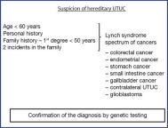
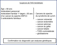
Specific UTUC risk factors | Aristolochic acid (AA) is the active principle of the aristolochiaceae family of herbaceous plants. Accidental ingestion or use in traditional pharmacopoeia is associated with a higher incidence of UTUC in the Balkans and on the Asian continent (Balkan Endemic Nephropathy, Chinese Herb Nephropathy) [6, 7]. A panel of experts has developed diagnostic criteria for AA nephropathy [8]. Two recent studies report that UTUC associated with AA are more common but have a better prognosis in women [9, 10]. Regular and prolonged consumption of phenacetin contained in various analgesic preparations has been questioned as a risk factor for UTUC from as early as 1965. The use of this molecule as an analgesic has been banned since the 1970s [2]. A particularly high incidence of UTUC (20 to 26.6% of all urothelial carcinomas) is also found on the south west coast of the island of Taiwan [11] associated with peripheral vasculitis called “black foot disease”, related to the concentration of arsenic in the water [12]. Lynch syndrome predisposes to several cancers transmitted by autosomal dominant inheritance. It is caused by the constitutional mutation of one of the genes of the DNA mismatch repair system (MLH1, MSH2, MSH6, PMS2). UTUC is the third most common in terms of localisation (about 5%) on the spectrum of Lynch Syndrome tumours, after colorectal and endometrial localisations [13]. The relative risk of developing a UTUC in case of Lynch syndrome ranges from 14 to 22. The MSH2 mutation is more commonly associated with the risk of UTUC [14]. Using the Amsterdam criteria, practitioners can consider the possibility of UTUC associated with Lynch syndrome (10–20% of UTUCs) but a positive diagnosis requires molecular confirmation [15] (Figure 1).
Figure 1. Clinical criteria for suspicion of hereditary UTUC.
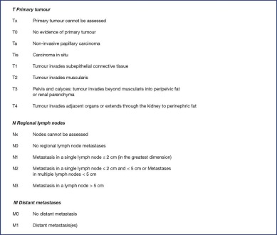
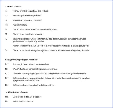
The WHO 2016 histological classification and tumour grade for UTUC are identical to those for BC treated in another chapter of the guidelines [16]. These tumours are urothelial carcinomas in more than 95% of cases [17]. UTUCs are non-invasive lesions (papillary exophytic proliferation of the urothelium with three distinct entities defined by the WHO 2016 classification to reflect the variable potential for progression) or invasive lesions (lesion that cross the basement membrane and reach at least the lamina propria ). The presence of “variant” components is found in approximately 25% of cases and is a poor prognostic factor [18, 19]. The UIIC 2017 TNM classification of tumour infiltration is shown in Figure 2.
Figure 2. TNM UIIC 2017 classification of UTUC.
|
|
Diagnosis and evaluation of UTUC |
| | Imaging examination: CTU & MRU | CT urography (CTU) is the reference imaging modality for the diagnostic workup of UTUC in patients with creatinine clearance > 30 ml/min [20, 21, 22]. It examines the entire urinary system through several acquisitions taken before and after the injection of contrast media and should include a study in the excretory phase of contrast medium elimination. The use of a protocol with furosemide (Lasilix®) injection and a double injection of contrast media (split bolus) is recommended to improve the performance of the examination and reduce patient irradiation [23]. The effectiveness of CTU for the diagnosis of UTUC is high (>90%) with pooled Sensitivity = 92% and pooled Specificity = 95% [24, 25, 26, 27]. The diagnostic performance of CTU decreases with flat lesions or lesions smaller than 5 mm. Magnetic Resonance Urography (MRU) also makes it possible to study the entire urinary system and is an interesting alternative to CTU, especially if CTU is contraindicated [2028-31]
| | Endoscopic and histological assessment | Urinary cytology is used to analyse cells from natural desquamation of the urothelial lining of the urinary tract. Cytology is recommended in the diagnosis of UTUC, although it is less sensitive and specific than in BC, including high-grade lesions. It should ideally be performed in situ (selective, during an endoscopic examination) and before any contrast medium is injected [1732, 33]. Positive urinary cytology predicts high-grade tumour with a 56% sensitivity and infiltrating tumour with a 62% sensitivity [34]. Since December 2015, a new world classification for urine cytology has been published [35]. As with BC, Table 1 summarises the course of action to be taken in urology based on the results according to this classification. At present, no identification of biological markers is recommended systematically in UTUC.
|
|
Cystoscopy and retrograde pyelography | Cystoscopy is recommended as part of the routine evaluation of UTUC because of the risk of synchronous bladder injury in 8 to 13% of cases [32, 33]. A normal cystoscopic examination associated with suspicious cytology for high-grade carcinoma is suggestive of high-grade UTUC. Retrograde pyelography (RP) can be performed in an emergency setting or when optimal upper urinary tract imaging has not been achieved. Under optimal conditions, PR has a sensitivity of 97% and a specificity of 93% for the detection of UTUC [24,33].
|
|
Flexible ureterorenoscopy (URS) | URS has improved the preoperative evaluation of UTUC by allowing macroscopic exploration of more than 95% of the entire upper system (including the lower calyces) [32, 33]. It provides information on the macroscopic aspect, the number of lesions (7 to 23% multifocal lesions) and allows the performance of biopsies and in situ cytologies [2]. Biopsies establish the diagnosis of urothelial carcinoma with a sensitivity of 89 to 95% [32]. The reliability of a biopsy in predicting the tumour stage is low with a high rate of underestimation (45% of Ta lesions are actually infiltrating tumours) [32, 33]. On the other hand, the biopsy grade is consistent with the final tumour grade in 69 to 91% of the cases. There is also an association between biopsy grade and final tumour stage. Biopsies revealing low-grade lesions correspond to a non-invasive tumour (≤ pT1) in 68 to 100% of the cases. Biopsies that identify high-grade lesions correspond to an infiltrating tumour (≥ pT2) in 62 to 100% of the cases [32,36]. URS should be systematically performed: • | in case of a doubtful diagnosis: ◦ | positive urinary cytologies without lesions established by cystoscopy and cross-sectional imaging.
| ◦ | benign lesion suspected on imaging (cystic ureteritis, fibroepithelial polyp, etc.)
|
| • | when conservative treatment is being considered.
|
However, the value of diagnostic URS has not been demonstrated in infiltrating or locally advanced lesions for which the reference treatment remains nephroureterectomy. In addition, recent data from the literature including 2 meta-analyses [37, 38, 39, 40] showed an increased risk of bladder cancer recurrence after RNU when patients had a diagnostic URS. The use of a catheter during the URS procedure to assess UTUC remains controversial, given the risk of seeding and the risk of not viewing lesions masked by the catheter. It is recommended to use low pressures when exploring the UUT. Detection sensitivity can be improved by various optical techniques. Oral 5-ALA used in a prospective study of 31 UTUCs improved the detection of carcinoma lesions in situ [41]. Other detection tools (Narrow Band Imaging [NBI], Storz Professional Image Enhancement System [SPIES]) are being developed [42]. In the event of an infiltrating lesion, a chest CT completes the staging of distant metastasis. The classic localisations for metastasis are the lungs (52%), liver (33%) and bone (26%) [32]. If there are clinical signs suggestive of metastatic lesions, a bone scintigraphy or brain scan may be performed [43]. A PET scan (with FDG or labelled Choline) is not recommended as part of the staging of the UTUC (#tabr1) [33,44].
| | Stratification of the risk of progression and prognostic factors |
|
|
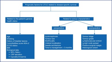
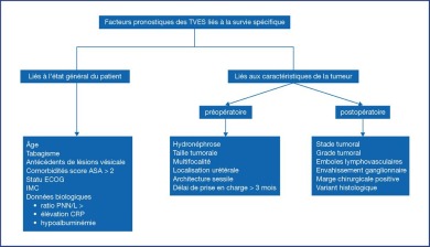
Preoperative prognostic factors | Several preoperative factors are associated with the risk of tumour progression (Figure 3). These are factors related to the patient (age, smoking, history of bladder lesions, altered general health, sarcopenia, biological criteria) or to the characteristics of the tumour (tumour size, multifocality, ureteral localisation, sessile architecture, hydronephrosis) [45, 46, 47, 48, 49, 50].
Figure 3. Prognostic factors for UTUC.
Time to surgical management > 3 months is associated with a poorer prognosis [51]. Preoperative nomograms based on these variables can predict the risk of locally advanced disease with an accuracy ranging from 77% to 82% [4852, 53].
|
|
Stratification of the risk of progression | Preoperative stratification of the risk of progression of UTUC is essential if conservative treatment is to be envisaged for low-risk tumours and radical treatment for high-risk tumours [54, 55] (Table 2): • | Low-risk UTUC: low grade on biopsy, and low grade cytology, and non-invasive appearance on imaging, and unifocal disease and size < 2 cm with the possibility of complete conservative treatment.
| • | High-risk UTUC: high grade on biopsy, or high grade cytology, or invasive appearance on imaging, or presence of hydronephrosis, or multifocal disease, or potentially incomplete resection of conservative therapy. Active smoking as well as a history of bladder lesions expose to recurrence and lower disease-specific survival.
|
|
|
Postoperative prognostic factors |
|
|
Disease-specific survival |
The prognostic factors after radical treatment are essentially pathological [5456, 57]. • | The stage and grade of the tumour are the main factors. The prognosis for UTUC is poor when they invade the muscle wall with a specific survival at 5 years of < 50% for pT2/pT3 and < 10% for pT4. On the contrary, disease-specific survival for tumours < pT2 is very good, almost 90% [ 52, 58]. | • | Tumour emboli with lymphovascular invasion are a prerequisite for lymph node spread.
| • | Lymph node spread is a negative factor for survival (in case of pN+ status; specific survival at 5 years = 35–40%).
| • | The presence of a positive surgical margin is an unfavourable element after RNU. The pathologist should examine this factor in relation to the ureteral section, the bladder neck and the tissue surrounding the tumour.
| • | The presence of a sarcomatoid or micropapillary variant is a factor of poor prognosis [ 19]. | • | Other negative pathological prognostic factors: presence of concomitant CIS, presence of tumour necrosis, multifocality and ureteral localisation.
| • | Molecular markers: patients with high intratumoral microsatellite instability (High MSI status) have better specific survival.
|
No other marker is used in daily clinical practice at present. Several nomograms to predict specific survival from these postoperative data have been proposed [54,59]. The nomogram by the French collaborative group has been externally validated [60, 61].
|
|
Bladder cancer recurrence |
Factors for bladder cancer recurrence after radical treatment were confirmed in a meta-analysis [4]. Three categories of risk factors were identified: • | patient-dependent factors (male, history of bladder lesion, preoperative chronic renal failure)
| • | tumour-dependent factors (positive preoperative urinary cytology, ureteral localisation, stage ≥ pT2, necrosis)
| • | treatment-dependent factors (laparoscopic approach, extravesical bladder neck approach, positive surgical margin and a diagnostic URS before RNU) [ 37, 38, 39, 40]. |
A nomogram incorporating these risk factors has been developed to predict the risk of intravesical recurrence but has not yet been externally validated [62].
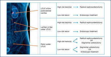
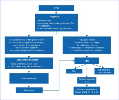
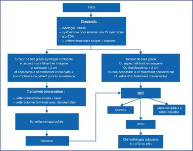
| | Conservative treatment of UTUC prevents the morbidity of radical therapy, including impaired renal function, without compromising the oncological outcomes [63]. Therefore, it should be routinely considered for low-risk tumours, even in the absence of contralateral kidney abnormalities. Figure 4. Management algorithm for localised UTUC based on localisation. Figure 5. Management algorithm for localised UTUC based on risk stratification. Flexible ureteroscopy allows exploration of the entire upper urinary tract and should be chosen over rigid ureteroscopy. A laser generator and biopsy equipment are required for treatment [33]. Patients should be warned of the risk of recurrence and should agree to close surveillance [64]. Some authors recommend that there should be a systematic “second look” within 60 days [65]. The only criterion associated with the risk of progression is the presence of a high tumour grade, irrespective of tumour size and the uni- or multifocal nature, provided that vaporisation of the lesion is complete [66]. The percutaneous approach is hardly ever used. It can be considered in case of lesions of the lower calyx inaccessible to treatment by URS [33,64]. Segmental ureterectomy allows the tumour to be removed en bloc with precise determination of the stage and histological grade. Lymphadenectomy can be performed at the same time. Distal ureterectomy with ureterovesical reimplantation for lower ureteral lesions has a better success rate than segmental resection of the iliac or lumbar ureter [67]. Partial surgery of the pyelocaliceal cavities is difficult and hardly ever performed in practice.
|
|
Instillations of topical adjuvant agents | Instillations of BCG or mitomycin C in the upper urinary tract are feasible for the treatment of ICIS or as adjuvant therapy after conservative treatment of low-risk papillary tumours. Anterograde administration by nephrostomy appears preferable to the retrograde approach by a ureteral catheter, which can be dangerous because of the risk of obstruction. Vesicoureteral reflux obtained by a double J stent has also been described but was not reproducible in all patients [68]. The effectiveness of these instillations is yet to be demonstrated [69]. A new topical hydrogel treatment containing mitomycin has been shown to be effective for the chemoablation of low-grade UTUC (#tabr2 and appendix 1) [70].
|
|
Radical nephroureterectomy |
Open radical nephroureterectomy (RNU) with excision of the peri-meatic bladder cuff is the standard surgical treatment for UTUC, regardless of localisation [33,71]. Primary ligation of the ureter below the level of the tumour reduces the risk of bladder cancer recurrence [72]. Oncological outcomes of the minimally invasive laparoscopic approach appear to be equivalent with less morbidity [7173, 74]. Certain oncological principles should be respected during laparoscopy: • | no opening of the urinary tract
| • | no fragmentation of the tumour
| • | use of an endoscopic pouch for the extraction of the surgical specimen in one piece (en bloc)
| • | excision of the kidney, ureter and bladder cuff en bloc
|
However, in case of locally advanced tumours (cT3/T4 and/or cN+), the laparoscopic approach is contraindicated because the oncological outcomes are poorer than with the open approach [73, 74]. The robot-assisted laparoscopic approach seems to provide comparable results to the other approaches [75, 76].
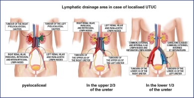
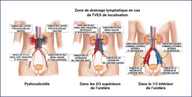
| | Excision of the intramural ureter and bladder cuff | Excision of the intramural portion of the ureter along with the ureteral meatus is recommended because of the high risk of local recurrence if a ureteral stump remains [4]. Several surgical techniques have been described: endoscopic resection, extra-vesical dissection, trans-vesical dissection [77]. The extra-vesical technique, especially with laparoscopy, appears to be associated with a higher rate of bladder cancer recurrence [4,62]. Lymphatic drainage areas of the upper urinary tract are not clearly defined. However, lymphadenectomy in combination with RNU enables better staging, guides therapeutic management (adjuvant chemotherapy) and could improve survival by reducing the risk of recurrence for tumours ≥ pT2 [7178, 79-80]. The extent of lymphadenectomy could have a greater prognostic value than the total number of lymph nodes removed [79]. Depending on the localisation of the tumour, lymphadenectomy may include primary, external and internal iliac and ileo-obturator areas in case of UTUC of the pelvic ureter, and in case of UTUC of the iliac/lumbar or pyelocaliceal ureter, the retroperitoneal area is included (internal limits on the right are the inferior vena cava and on the left, the aorta) (Figure 6). Figure 6. Proposal of a lymphadenectomy zone according to the localisation of the tumour and the lymphatic drainage.
|
|
Distal ureterectomy with reimplantation | In case of isolated lesions of the lower ureter, distal ureterectomy with ureterovesical reimplantation allows the kidney to be preserved with good oncological control and the possibility of lymphadenectomy [6381, 82]. However, given the low level of evidence, this strategy can only be proposed to select high-risk UTUC cases.
|
|
Essential conservative treatments | In case of a single functional or anatomical kidney, bilateral tumours or co-morbidities that prevent radical treatment, conservative treatment of necessity may be considered for high-risk UTUC. Endoscopic treatment is associated with a higher rate of progression for high-grade tumours [63].
|
|
Adjuvant intravesical instillations |
The bladder cancer recurrence rate after RNU is between 20 and 40% [4]. Two prospective randomised trials have demonstrated the benefit of early postoperative instillation of intravesical chemotherapy (IVCT) (mitomycin C or pirarubicin) in reducing the risk of bladder cancer recurrence (11% absolute risk reduction) [83, 84]. After conservative treatment, early postoperative IVCT may also be beneficial [85].
| | Neoadjuvant/adjuvant therapies | Neoadjuvant platinum-based chemotherapy has the theoretical advantage of the possibility of being administered to the majority of patients, due to the risk of impaired renal function after RNU [86]. Several retrospective studies have shown a benefit of neoadjuvant chemotherapy in terms of pathological response after RNU, recurrence-free survival and disease-specific survival (#tabr3) [87, 88]. Adjuvant chemotherapy allows the selection of patients on the basis of pathological findings. The randomised trial POUT showed that platinum-based chemotherapy started within 90 days of RNU with lymphadenectomy significantly improved disease-free survival in patients with ≥ pT2 or pN+ tumours [89]. Adjuvant radiotherapy might improve local disease control, but the effects on survival are controversial and it is currently not recommended [90]. For metastatic disease, platinum-based chemotherapy is the gold standard, with results comparable to those for bladder tumours [91]. Immunotherapy has shown promising first-line results in cisplatin ineligible patients [92, 93] and several trials are under way for 2nd line treatment. Local treatment by surgery or radiotherapy is not indicated except for palliative care of symptoms. It can be discussed in case of a response to induction chemotherapy and several retrospective studies have shown a benefit in select patients [94, 95]. Post-treatment surveillance of UTUC is required in order to detect bladder cancer recurrence, locally or remotely [33,96]. After RNU, the risk of local recurrence is low, whereas the risk of metastatic recurrence is directly dependent on the prognostic factors. This risk of recurrence changes over time and tends to decrease during follow-up [97]. Surveillance is based on urinary cytology, cystoscopy and a CT urography supplemented by a chest CT scan if the lesion is invasive. The frequency of surveillance after radical treatment is reported in #tabr4.
| | After conservative treatment | After conservative treatment, the ipsilateral urinary tract requires special monitoring because of the high risk of local recurrence. A «second look» by ureteroscopy at 6 weeks from the laser treatment is recommended by some experts [65]. Surveillance is based on urinary and in situ cytology, cystoscopy, ureteroscopy and CTU. The frequency of surveillance should be increased.
|
|
Épidémiologie - Facteurs de risque | On estime que 5 % des carcinomes urothéliaux sont des TVES et l’incidence est approximativement de 1/100 000 habitants/an [1, 2]. Bien que les TVES aient le même aspect anatomopathologique que les tumeurs de la vessie (TV), elles présentent un certain nombre de particularités sur les plans épidémiologique, clinique et pronostique. Le pic d’incidence des TVES est compris entre 70 et 90 ans, avec un ratio homme/femme proche de 2 pour 1 [2]. Les TVES sont diagnostiquées à un stade invasif dans 60 % des cas [2] et la maladie est métastatique d’emblée dans 7 % des cas [3]. La lésion est pyélocalicielle dans 60 % des cas, urétérale dans 30 % des cas et multifocale dans 10 % des cas [2]. Un antécédent de lésion vésicale est retrouvé chez 15 à 30 % des patients. Les récidives vésicales après traitement radical d’une TVES sont fréquentes (30 à 50 %) alors que l’incidence des TVES après une TV est faible (2 à 5 %) [4].
| | Facteurs de risque et facteurs génétiques |
|
|
Facteurs de risque communs avec les TV | Le tabac constitue un facteur de risque majeur (augmentation du risque relatif de 2,5 à 7). Ce risque est modulé par l’intensité de la consommation tabagique et décroît après l’arrêt de l’exposition tabagique. La poursuite de la consommation après diagnostic constituerait un facteur pronostique défavorable [2,5]. L’exposition professionnelle aux amines aromatiques, hydrocarbures aromatiques polycycliques (HAP) et solvants chlorés constitue également un facteur de risque de TVES [2]. L’exposition chronique à l’acroléine (métabolite actif du cyclophosphamide) est un facteur de risque de TVES [2]. Ce type de chimiothérapie est à éviter en cas d’antécédent de carcinome urothélial ou doit être associé à la prise d’un protecteur urothélial (MESNA ou sodium 2-mercaptoethane sulfate). Les infections et les inflammations chroniques (maladie lithiasique) constituent des facteurs de risque de TVES de type épidermoïde ou adénocarcinomateux [2].
|
|
Facteurs de risque propres aux TVES | L’acide aristolochique (AA) est le principe actif des plantes herbacées aristoloches. Son ingestion accidentelle ou son utilisation en pharmacopée traditionnelle est associée à une surincidence des TVES dans les Balkans et sur le continent asiatique (néphropathie des Balkans, néphropathie aux herbes chinoises) [6, 7]. Un panel d’experts a défini des critères diagnostiques de néphropathie aux AA [8]. Deux études récentes rapportent que les TVES associées à l’AA sont plus fréquentes mais de meilleur pronostic chez les femmes [9, 10]. La consommation régulière et prolongée de phénacétine contenue dans diverses préparations antalgiques a été mise en cause comme facteur de risque de TVES dès 1965. L’utilisation de cette molécule comme antalgique est interdite depuis les années 1970 [2]. Une incidence particulièrement élevée de TVES (20 à 26,6 % de l’ensemble des carcinomes urothéliaux) est également retrouvée sur la côte sud-ouest de l’île de Taiwan [11], associée à une vascularite périphérique appelée « maladie du pied noir » ou blackfoot disease, en rapport avec la concentration d’arsenic dans l’eau [12]. Le syndrome de Lynch est un syndrome de prédisposition à plusieurs cancers, transmis sur un mode autosomique dominant. Il résulte de la mutation constitutionnelle d’un des gènes du système de réparation des mésappariements de l’ADN (MLH1, MSH2, MSH6, PMS2 ). Les TVES sont la troisième localisation la plus fréquente (environ 5 %) du spectre des tumeurs associées au syndrome de Lynch, après les localisations colorectales et endométriales [13]. Le risque relatif de développer une TVES en cas de syndrome de Lynch varie de 14 à 22. La mutation du MSH2 est plus souvent associée au risque de TVES [14]. Les critères d’Amsterdam permettent aux praticiens de suspecter les TVES associées à un syndrome de Lynch (10 à 20 % des TVES), mais le diagnostic positif nécessite une confirmation moléculaire (Figure 1) [15]. Figure 1. Critères cliniques de suspicion d’une TVES héréditaire. La classification histologique et le grade tumoral OMS 2016 des TVES sont identiques à ceux des TV traitées dans un autre chapitre des recommandations [16]. Ces tumeurs sont des carcinomes urothéliaux dans plus de 95 % des cas [17]. Les TVES sont des lésions non invasives (prolifération exophytique, papillaire de l’urothélium avec trois entités distinctes définies par la classification OMS 2016, afin de refléter le potentiel évolutif variable) ou des lésions invasives (lésion franchissant la membrane basale et atteignant la lamina propria au minimum). La présence de contingents « variants » est retrouvée dans environ 25 % des cas, et constitue un facteur de mauvais pronostic [18, 19]. La classification de l’infiltration tumorale TNM UIIC 2017 est exposée dans la Figure 2. Figure 2. Classification TNM UIIC 2017 des TVES.
|
|
Diagnostic et évaluation des TVES |
| | Bilan d’imagerie : uro-TDM, uro-IRM | L’uroscanner (uro-TDM) constitue la modalité d’imagerie de référence pour le bilan diagnostique des TVES chez les patients avec une clairance de créatinine > 30 ml/min [20, 21, 22]. Il étudie l’ensemble de l’appareil urinaire par plusieurs acquisitions réalisées avant et après injection de produit de contraste et comporte obligatoirement une étude à la phase excrétoire de l’élimination du produit de contraste. L’utilisation d’un protocole avec injection de furosémide (Lasilix®) et double injection de produit de contraste (split bolus ) est recommandée pour améliorer les performances de l’examen et diminuer l’irradiation des patients [23]. L’efficacité de l’uro-TDM pour le diagnostic des TVES est élevée (> 90 %) avec une sensibilité poolée = 92 % et une spécificité poolée = 95 % [24, 25, 26, 27]. Les performances diagnostiques de l’uro-TDM diminuent en cas de lésions planes ou inférieures à 5 mm. L’imagerie par résonance magnétique urinaire (uro-IRM) permet également d’étudier l’ensemble de l’appareil excrétoire urinaire et constitue une alternative intéressante à l’uro-TDM, notamment si celle-ci est contre-indiquée [2028-31].
| | Bilan endoscopique et histologique |
|
|
Cytologie - Marqueurs biologiques | La cytologie urinaire permet d’analyser des cellules provenant de la desquamation naturelle du revêtement urothélial des voies urinaires. La cytologie est recommandée dans le diagnostic des TVES bien qu’elle soit moins sensible et spécifique que dans les cas de TV, y compris pour des lésions de haut grade. Elle doit idéalement être réalisée in situ (sélective, au cours d’un examen endoscopique) et avant toute injection de produit de contraste [1732, 33]. Une cytologie urinaire positive prédit une tumeur de haut grade avec une sensibilité de 56 % et une tumeur infiltrante avec une sensibilité de 62 % [34]. Depuis décembre 2015, une nouvelle classification mondiale pour la cytologie urinaire a été publiée [35]. Comme pour les TV, le Tableau 1 résume la conduite à tenir en fonction des résultats de cette classification. Aucune recherche de marqueur biologique n’est aujourd’hui recommandée de manière systématique dans les TVES.
|
|
Cystoscopie et urétéropyélographie rétrograde | La réalisation d’une cystoscopie est recommandée dans le bilan systématique d’une TVES en raison d’un risque de lésion vésicale synchrone dans 8 à 13 % des cas [32, 33]. Un examen cystoscopique normal associé à des cytologies suspectes pour un carcinome de haut grade est évocateur d’une TVES de haut grade. L’urétéropyélographie rétrograde (UPR) peut être réalisée dans un contexte d’urgence ou lorsqu’une imagerie optimale du haut appareil urinaire n’a pas pu être effectuée. Dans des conditions optimales, l’UPR a une sensibilité de 97 % et une spécificité de 93 % pour la détection des TVES [24,33].
|
|
Urétérorénoscopie souple (URSS) | L’URSS a amélioré l’évaluation préopératoire des TVES en permettant l’exploration macroscopique de plus 95 % de l’ensemble du haut appareil (y compris des calices inférieurs) [32, 33]. Elle renseigne sur l’aspect macroscopique, le nombre de lésions (7 à 23 % de lésions multifocales) et permet la réalisation de biopsies et de cytologies in situ [2]. Les biopsies établissent le diagnostic de carcinome urothélial avec une sensibilité de 89 à 95 % [32]. La fiabilité de la biopsie dans la prédiction du stade tumoral est faible avec un taux de sous-évaluation important (45 % des lésions Ta sont en réalité des tumeurs infiltrantes) [32, 33]. En revanche, le grade biopsique est concordant avec le grade tumoral définitif dans 69 à 91 % des cas. Il existe également une association entre le grade biopsique et le stade tumoral définitif. Les biopsies révélant des lésions de bas grade correspondent à une tumeur non infiltrante (≤ pT1) dans 68 à 100 % ; les biopsies trouvant du haut grade correspondent à une tumeur infiltrante (≥ pT2) dans 62 à 100 % des cas [32,36]. L’URSS doit être systématiquement réalisée: • | en cas de doute diagnostique: ◦ | cytologies urinaires positives sans lésion objectivée en cystoscopie et imagerie en coupe,
| ◦ | lésion bénigne suspectée en imagerie (urétérite kystique, polype fibroépithélial…);
|
| • | lorsqu’un traitement conservateur est envisagé.
|
En revanche, l’intérêt de l’URSS diagnostique n’a pas été démontré en cas de lésion infiltrante ou localement avancée pour laquelle le traitement de référence reste la néphrourétérectomie. De plus, les données récentes de la littérature dont deux méta-analyses [37, 38, 39, 40] ont montré une augmentation du risque de récidive vésicale après NUT lorsque les patients avaient une URSS diagnostique. L’utilisation d’une gaine d’accès lors du geste d’URSS pour bilan d’une TVES reste controversée, compte tenu du risque d’essaimage et du risque de ne pas visualiser des lésions masquées par la gaine. Il est recommandé d’utiliser de faibles pressions lors de l’exploration de la voie excrétrice urinaire supérieure (VES). La sensibilité de détection peut être améliorée par différentes techniques optiques. Le 5-ALA utilisé par voie orale dans une étude prospective de 31 TVES permettait d’améliorer la détection des lésions de carcinome in situ (CIS) [41]. D’autres outils de détection (Narrow Band Imaging [NBI], Storz Professional Image Enhancement System [SPIES]) sont en cours de développement [42]. En cas de lésion infiltrante, un scanner thoracique complète le bilan d’extension à distance. Les localisations métastatiques classiques sont pulmonaires (52 %), hépatiques (33 %) et osseuses (26 %) [32]. En cas de signes cliniques évocateurs de lésions métastatiques, une scintigraphie osseuse ou un scanner cérébral peuvent être réalisés [43]. La réalisation d’un TEP-scanner (au FDG ou à la choline marquée) n’est pas recommandée dans le cadre du bilan d’extension des TVES (#ntabr1) [33,44].
| | Stratification du risque évolutif et facteurs pronostiques |
|
|
Facteurs pronostiques préopératoires | Plusieurs facteurs préopératoires sont associés au risque évolutif de la tumeur (Figure 3). Il s’agit de facteurs liés au patient (âge, tabagisme, antécédents de lésions vésicales, altération de l’état général, sarcopénie, critères biologiques) ou aux caractéristiques de la tumeur (taille tumorale, multifocalité, localisation urétérale, architecture sessile, hydronéphrose) [45, 46, 47, 48, 49, 50]. Figure 3. Facteurs pronostiques des TVES. Un délai de prise en charge chirurgicale > 3 mois est associé à un moins bon pronostic [51]. Des nomogrammes préopératoires construits à partir de ces variables permettent de prédire le risque de maladie localement avancée avec une précision allant de 77 à 82 % [4852, 53].
|
|
Stratification du risque évolutif | La stratification préopératoire du risque évolutif des TVES est essentielle afin d’envisager un traitement conservateur pour les tumeurs de bas risque et un traitement radical pour les tumeurs de haut risque [54, 55] (Tableau 2): • | TVES à bas risque : faible grade sur biopsie, et faible grade cytologique, et aspect non infiltrant en imagerie, et maladie unifocale et taille < 2 cm avec possibilité d’un traitement conservateur complet;
| • | TVES à haut risque : haut grade sur biopsie, ou haut grade cytologique, ou aspect infiltrant en imagerie, ou présence d’une hydronéphrose, ou maladie multifocale, ou résection potentiellement incomplète du traitement conservateur. Le tabagisme actif, de même que les antécédents de lésions vésicales exposent à des récidives et à une survie spécifique altérée.
|
|
|
Facteurs pronostiques postopératoires |
Les facteurs pronostiques après traitement radical sont essentiellement anatomopathologiques [5456, 57]. • | Le stade et le grade de la tumeur sont les facteurs principaux. Le pronostic des TVES est sombre lorsqu’elles infiltrent la paroi musculaire avec une survie spécifique à 5 ans < 50 % pour les pT2/pT3 et < 10 % pour les pT4. Au contraire, la survie spécifique des tumeurs < pT2 est très bonne, proche de 90 % [52,58].
| • | Les emboles lymphovasculaires tumoraux sont un préalable à l’envahissement ganglionnaire.
| • | L’envahissement ganglionnaire est un facteur péjoratif de survie (en cas de statut pN+ ; survie spécifique à 5 ans = 35 à 40 %).
| • | La présence d’une marge chirurgicale positive est un élément défavorable après NUT. Le pathologiste doit rapporter ce facteur au niveau de la section urétérale, de la collerette vésicale et du tissu au pourtour de la tumeur.
| • | La présence d’un variant sarcomatoïde ou micropapillaire est un facteur de mauvais pronostic [ 19]. | • | Autres facteurs pronostiques anatomopathologiques péjoratifs : présence de CIS concomitant, présence d’une nécrose tumorale, multifocalité et localisation urétérale.
| • | Marqueurs moléculaires : les patients avec une forte instabilité intratumorale des microsatellites (statut MSI High) ont une meilleure survie spécifique.
|
Aucun autre marqueur n’est aujourd’hui utilisé en pratique clinique quotidienne. Plusieurs nomogrammes permettant de prédire la survie spécifique à partir de ces données postopératoires ont été proposés [54, 59]. Le nomogramme du groupe collaboratif français a bénéficié d’une validation externe [60, 61].
Les facteurs de récidive vésicale après traitement radical ont été confirmés dans une méta-analyse [4]. Trois catégories de facteurs de risque ont été identifiées: • | facteurs dépendant du patient (sexe masculin, antécédent de lésion vésicale, insuffisance rénale chronique préopératoire) ;
| • | facteurs dépendant de la tumeur (cytologie urinaire positive préopératoire, localisation urétérale, stade ≥ pT2, nécrose) ;
| • | facteurs dépendant du traitement (approche laparoscopique, abord extravésical de la collerette vésicale, marge chirurgicale positive et réalisation d’une URSS diagnostique avant la NUT) [ 37, 38, 39, 40]. |
Un nomogramme intégrant ces facteurs de risque a été développé pour prédire le risque de récidive intravésicale, mais n’a pas bénéficié d’une validation externe à ce jour [62].
| |
| | TVES localisée à faible risque | Le traitement conservateur des TVES permet d’éviter la morbidité d’un traitement radical, notamment la dégradation de la fonction rénale, sans compromettre les résultats carcinologiques [63]. Par conséquent, il doit être systématiquement envisagé en cas de tumeur à faible risque, même en l’absence d’anomalie du rein controlatéral. Figure 4. Algorithme de prise en charge des TVES localisées en fonction de la localisation. Figure 5. Algorithme de prise en charge des TVES localisées en fonction de la stratification du risque. L’urétéroscopie souple permet d’explorer l’ensemble de la VES et doit être préférée à l’urétéroscopie rigide. Un générateur laser et du matériel pour biopsie sont nécessaires au traitement [33]. Les patients doivent être prévenus du risque de récidive et accepter une surveillance rapprochée [64]. Certains auteurs recommandent la réalisation systématique d’un « second look » dans les 60 jours [65]. Le seul critère associé au risque de progression est la présence de haut grade tumoral, indépendamment de la taille tumorale et du caractère uni- ou multifocal, sous réserve que la vaporisation de la lésion soit complète [66]. L’approche percutanée est très peu utilisée. Elle est envisageable en cas de lésion du calice inférieur, inaccessible au traitement par URSS [33,64].
|
|
Urétérectomie segmentaire | L’urétérectomie segmentaire permet l’exérèse en monobloc de la tumeur avec une détermination précise du stade et du grade histologique. Un curage ganglionnaire peut être réalisé dans le même temps. L’urétérectomie terminale avec réimplantation urétéro-vésicale pour lésion du bas uretère présente un meilleur taux de succès que la résection segmentaire de l’uretère iliaque ou lombaire [67]. La chirurgie partielle des cavités pyélocalicielles est difficile et très peu réalisée en pratique.
|
|
Instillations d’agents topiques adjuvants | Les instillations du bacille de Calmette et Guérin (BCG) ou mitomycine C dans la VES sont faisables, pour le traitement du CIS, ou en adjuvant après traitement conservateur de lésions papillaires à faible risque. L’administration antérograde par une néphrostomie semble préférable à la voie rétrograde par une sonde urétérale qui peut être dangereuse en raison du risque d’obstruction. Le reflux vésico-urétéral obtenu par une sonde JJ a également été décrit mais n’est pas reproductible chez tous les patients [68]. L’efficacité de ces instillations reste à démontrer [69]. Un nouveau traitement topique par hydrogel contenant de la mitomycine a montré son efficacité pour la chimio-ablation des TVES de bas grade (#ntabr2) (annexe 1) [70].
| | TVES localisée à haut risque |
|
|
Traitement radical par néphrourétérectomie totale |
La NUT par voie ouverte avec excision d’une collerette vésicale périméatique est le traitement chirurgical de référence des TVES, quelle que soit la localisation [33,71]. La ligature première de l’uretère sous le niveau de la tumeur permet de diminuer le risque de récidive vésicale [72]. Les résultats oncologiques de la voie mini-invasive par laparoscopie semblent équivalents avec une morbidité moindre [7173, 74]. Certains principes oncologiques doivent être respectés lors de la laparoscopie: • | pas d’ouverture de la voie excrétrice ;
| • | pas de morcellation de la tumeur ;
| • | utilisation d’un sac endoscopique pour l’extraction de la pièce opératoire en monobloc ;
| • | exérèse du rein, de l’uretère et de la collerette vésicale en bloc.
|
Cependant, en cas de tumeur localement avancée (cT3/ T4 et/ou cN+), la voie laparoscopique est contre-indiquée car les résultats carcinologiques sont moins bons que la voie ouverte [73, 74]. La voie laparoscopique robot-assistée semble donner des résultats comparables aux autres voies d’abord [75, 76].
|
|
Exérèse de l’uretère intramural et de la collerette vésicale | L’exérèse de la portion intramurale de l’uretère emportant le méat urétéral est recommandée en raison du risque important de récidive locale en cas de persistance d’un moignon urétéral [4]. Plusieurs techniques chirurgicales ont été décrites : résection endoscopique, dissection extravésicale, dissection transvésicale [77]. La technique extravésicale, notamment en laparoscopie, semble associée à un taux plus élevé de récidive vésicale [4,62]. Les aires de drainage lymphatique de la VES ne sont pas clairement définies. Cependant, le curage ganglionnaire associé à la NUT permet une meilleure stadification, guide la prise en charge thérapeutique (chimiothérapie adjuvante) et pourrait améliorer la survie en diminuant le risque de récidive pour les tumeurs ≥ pT2 [7178, 79-80]. L’étendue du curage ganglionnaire aurait une valeur pronostique plus importante que le nombre total de ganglions prélevés [79]. En fonction de la localisation tumorale, le curage peut inclure un curage iliaque primitif, externe, interne et ilio-obturateur en cas de TVES de l’uretère pelvien ; un curage rétropéritonéal en cas de TVES de l’uretère iliaque/lombaire ou pyélocalicielle (limites internes à droite la veine cave inférieure et à gauche l’aorte) (Figure 6). Figure 6. Proposition de zone de curage en fonction de la localisation tumorale et de son drainage lymphatique.
|
|
Urétérectomie terminale avec réimplantation | En cas de lésion isolée du bas uretère, l’urétérectomie terminale avec réimplantation urétéro-vésicale permet de conserver le rein avec un bon contrôle carcinologique et la possibilité de réaliser un curage ganglionnaire [6381, 82]. Cependant, compte tenu du faible niveau de preuve, cette stratégie ne peut être proposée qu’à des cas sélectionnés de TVES à haut risque.
|
|
Cas particulier des traitements conservateurs impératifs | En cas de rein unique fonctionnel ou anatomique, de tumeurs bilatérales ou de comorbidités empêchant un traitement radical, un traitement conservateur de nécessité peut être envisagé pour une TVES à haut risque. Le traitement endoscopique est associé à un taux de progression plus important pour les tumeurs de haut grade [63].
|
|
Traitements périopératoires |
|
|
Instillations intravésicales adjuvantes |
Le taux de récidive vésicale après NUT est compris entre 20 et 40 % [4]. Deux essais prospectifs randomisés ont démontré le bénéfice d’une instillation postopératoire précoce (IPOP) de chimiothérapie intravésicale (mitomycine C ou pirarubicine) pour la diminution du risque de récidive vésicale (diminution absolue du risque de 11 %) [83, 84]. Après un traitement conservateur, l’IPOP pourrait également être bénéfique [85].
|
|
Traitements néo-adjuvants/adjuvants |
La chimiothérapie néo-adjuvante à base de sels de platine présente l’avantage théorique de pouvoir être administrée à un plus grand nombre de patients, en raison du risque de dégradation de la fonction rénale après NUT [86]. Plusieurs études rétrospectives ont montré un bénéfice de la chimiothérapie néo-adjuvante en termes de réponse pathologique après NUT, de survie sans récidive et de survie spécifique (#ntabr3) [87, 88]. La chimiothérapie adjuvante permet de sélectionner les patients sur la base des résultats anatomopa-thologiques. L’essai randomisé POUT a montré qu’une chimiothérapie à base de sels de platine débutée dans les 90 jours suivant la NUT avec curage améliorait significativement la survie sans récidive en cas de tumeur ≥ pT2 ou pN+ [89]. La radiothérapie adjuvante pourrait améliorer le contrôle local de la maladie, mais ses effets sur la survie sont controversés et elle est actuellement non recommandée [90]. En cas de maladie métastatique, la chimiothérapie à base de sels de platine est le traitement de référence, avec des résultats comparables aux TV [91]. L’immunothérapie a montré des résultats prometteurs en première ligne chez les patients inéligibles au cisplatine et plusieurs essais sont en cours en deuxième ligne [92, 93]. Le traitement local par chirurgie ou radiothérapie n’est pas indiqué en dehors du contexte palliatif symptomatique. Il peut être discuté en cas de réponse à une chimiothérapie d’induction et plusieurs études rétrospectives ont montré un bénéfice chez des patients sélectionnés [94, 95]. La surveillance après traitement d’une TVES est nécessaire afin de détecter une récidive vésicale, locale, ou à distance [33,96]. Après NUT, le risque de récidive locale est faible alors que le risque de récidive métastatique dépend directement des facteurs pronostiques. Ce risque de récidive évolue au fur et à mesure du temps et tend à décroître pendant le suivi [97]. La surveillance est basée sur la cytologie urinaire, la cystoscopie et un uroscanner complété par un scanner thoracique si la lésion est infiltrante. Le rythme de la surveillance après traitement radical est rapporté dans le #ntabr4.
| | Après traitement conservateur | Après traitement conservateur, la voie excrétrice homolatérale nécessite un suivi particulier du fait du risque important de récidive locale. Un second look par urétéroscopie à 6 semaines du traitement laser est recommandé par certains experts [65]. La surveillance repose sur la cytologie urinaire et in situ, la cystoscopie, l’urétéroscopie et l’uro-TDM. Le rythme de surveillance doit être plus rapproché.
|
|
Déclaration de liens d’intérêts | Morgan Rouprêt : honoraires board Ambu, Bayer, Janssen, Roche, MSD, Fidia, Ipsen, Astellas, Arquer, Cepheid, AstraZeneca, Ferring, Oncodiag. Géraldine Pignot : honoraires board Janssen, Bouchara-Recordati, BMS, Roche, Arquer, Astra-Zeneca, Astellas, Bayer. Alexandra Masson-Lecomte : honoraires board Ipsen, Janssen, AstraZeneca, Ambu. Eva Compérat : honoraires board Janssen, AstraZeneca, MSD. François Audenet : honoraires board Nucleix ; conferences Ferring, Ipsen, Janssen. Mathieu Roumiguié : honoraires board Janssen, Roche, Ipsen, Astellas, Arquer, AstraZeneca, Ferring, Oncodiag, Pierre Fabre. Nadine Houédé : honoraires board Astellas, AstraZeneca, Bayer, BMS, Ferring, Ipsen, Janssen, MSD, Novartis, Pfizer, Schugai. Stéphane Larré : honoraires board Nucleix, Sanofi. Serge Brunelle : pas de conflit. Evanguelos Xylinas : honoraires board AstraZeneca, Ferring, Janssen. Yann Neuzillet : honoraires board Astellas, AstraZeneca, BMS, Bouchara-Recordati, Ipsen, MSD et Sanofi Aventis. Arnaud Méjean : honoraires board Pfizer, Ipsen, Astellas, Ferring, Janssen, Novartis, GSK, BMS, Roche, Pierre Fabre ; intérêt institutionnel AFU, CCAFU, APHP, université de Paris, ARTuR.
|
|
Annexe 1. Argumentaire complémentaire |
| | Facteurs de risque de récidive vésicale après NUT | Une méta-analyse incluant 8 275 patients a montré un taux de récidive vésicale de 29 % avec un délai médian de 22,2 mois après NUT [Seisen T, Granger B, Colin P, Léon P, Utard G, Renard-Penna R, et al. A Systematic Review and Meta-analysis of Clinicopathologic Factors Linked to Intravesical Recurrence After Radical Nephroureterectomy to Treat Upper Tract Urothelial Carcinoma. Eur Urol 2015;67:1122–33 ]. Plusieurs facteurs de risque ont été identifiés : facteurs liés au patient (sexe masculin [HR 1.37 ; p < 0,001], antécédent de tumeur de vessie [HR 1,96 ; p < 0,001], insuffisance rénale chronique [HR 1,87 ; p = 0,002]), facteurs liés à la tumeur (cytologie positive [HR 1,56 ; p < 0,001], localisation urétérale [HR 1,27 ; p < 0,001], multifocalité [HR 1,61 ; p = 0,002], tumeur infiltrante [HR 1.38 ; p < 0,001], présence de nécrose [HR 2,17 ; p = 0,02]), facteurs liés au traitement (voie laparoscopique [HR 1,62 ; p = 0,003], collerette vésicale par voie extravésicale [HR 1,22 ; p = 0,02], marges chirurgicales positives [HR 1,90 ; p = 0,004]). L’analyse moléculaire des récidives vésicales après NUT est en faveur d’une origine clonale par dissémination intraluminale avec implantation secondaire [Audenet F, Isharwal S, Cha EK, Donoghue MTA, Drill EN, Ostrovnaya I, et al. Clonal Relatedness and Mutational Differences between Upper Tract and Bladder Urothelial Carcinoma. Clin Cancer Res 2019;25:967–76 ]. Plusieurs études rétrospectives ont évalué le traitement conservateur dans les TVES. Une méta-analyse publiée en 2016 a inclus 7 études pour un total de 547 patients traités de manière conservatrice et 1 376 patients traités par NUT [Seisen T, Peyronnet B, Dominguez-Escrig JL, Bruins HM, Yuan CY, Babjuk M, et al. Oncologic Outcomes of Kidney-sparing Surgery Versus Radical Nephroureterectomy for Upper Tract Urothelial Carcinoma: A Systematic Review by the EAU Non-muscle Invasive Bladder Cancer Guidelines Panel. Eur Urol 2016;70:1052–68 ]. Malgré un biais de sélection, il n’existait pas de différence significative entre urétérectomie segmentaire et NUT. Concernant le traitement endoscopique, seuls les patients avec une tumeur de bas grade et non infiltrante avaient une survie spécifique similaire à la NUT, malgré un risque plus élevé de récidive locale. Une étude rétrospective monocentrique incluant 92 patients avec une TVES traitée de manière conservatrice par urétéroscopie souple et photovaporisation laser a montré en analyse multivariée, que seul le grade tumoral était significativement associé au risque de progression (HR = 5,16 ; IC 95 % : 1,19–22,26 ; p = 0,03) [Villa L, Haddad M, Capitanio U, Somani BK, Cloutier J, Doizi S, et al. Which Patients with Upper Tract Urothelial Carcinoma Can be Safely Treated with Flexible Ureteroscopy with Holmium:YAG Laser Photoablation? Long-Term Results from a High Volume Institution. J Urol 2018;199:66–73 ]. La multifocalité ou la taille tumorale supérieure à 1 cm n’étaient pas des facteurs prédictifs du risque de progression. Une autre méta-analyse publiée en 2020 incluant 4 797 patients a montré que le traitement par urétérectomie segmentaire était associé à un risque de récidive plus élevé (HR 0,64 ; IC 95 % 0,43–0,95 ; p = 0,03), sans impact sur la survie spécifique ou la survie sans métastase, mais avec un meilleur débit de filtration glomérulaire [Veccia A, Antonelli A, Checcucci E, Falagario U, Carrieri G, Guruli G, et al. Segmental Ureterectomy for Upper Tract Urothelial Carcinoma: A Systematic Review and Metaanalysis of Comparative Studies. Clin Genitourin Cancer 2020;18:e10–20 ].
| | Instillations d’agents topiques adjuvants | Deux essais prospectifs randomisés ont démontré le bénéfice d’une instillation postopératoire précoce de chimiothérapie intravésicale pour la diminution du risque de récidive vésicale. Le premier essai publié en 2011 a randomisé 144 patients dans le bras mitomycine C (MMC) (40 mg dans 40 ml de sérum physiologique) administrée en postopératoire précoce et 140 patients dans le bras contrôle [O’Brien T, Ray E, Singh R, Coker B, Beard R. British Association of Urological Surgeons Section of Oncology. Prevention of bladder tumours after nephroureterectomy for primary upper urinary tract urothelial carcinoma: a prospective, multicentre, randomised clinical trial of a single postoperative intravesical dose of mitomycin C (the ODMIT-C Trial). Eur Urol 2011;60:703–10 ]. L’analyse en intention de traiter a montré un taux de récidive vésical à 1 an de 17 % dans le bras MMC versus 27 % dans le bras contrôle (p = 0,03). Le deuxième essai publié en 2013 a inclus 77 patients : 39 dans le bras traité par instillation de pirarubicine (30 mg dans 30 ml de sérum physiologique) administrée dans les 48 heures suivant la NUT et 38 patients dans le bras contrôle [Ito A, Shintaku I, Satoh M, Ioritani N, Aizawa M, Tochigi T, et al. Prospective randomized phase II trial of a single early intravesical instillation of pirarubicin (THP) in the prevention of bladder recurrence after nephroureterectomy for upper urinary tract urothelial carcinoma: the THP Monotherapy Study Group Trial. J Clin Oncol 2013;31:1422–7 ]. L’analyse en intention de traiter a montré un taux de récidive vésicale à 1 et 2 ans de 16,9 % et 16,9 % dans le bras THP versus 31,8 % et 42,2 % dans le bras contrôle. L’essai randomisé POUT, publié en 2020, a randomisé 261 patients avec une TVES classée pT2–4 N0–3 M0 ou pN+ M0 après NUT : 132 patients ont reçu 4 cycles de chimiothérapie adjuvante à base de cisplatine ou de carboplatine si DFG < 50 ml/min, débutée dans les 90 jours postopératoires et 129 dans le bras surveillance seule [Birtle A, Johnson M, Chester J, Jones R, Dolling D, et al. Adjuvant chemotherapy in upper tract urothelial carcinoma (the POUT trial): a phase 3, open-label, randomised controlled trial. Lancet ;395:1268–77 ]. Après un suivi médian de 30,8 mois, la chimiothérapie adjuvante était associée à une amélioration de la survie sans récidive (HR 0,45, IC 95 % 0,30–0,68 ; p = 0,0001 ). Le taux de survie sans récidive à 3 ans était de 71 % dans le bras chimiothérapie versus 46 % dans le bras contrôle. 44 % des patients traités par chimiothérapie ont présenté des effets indésirables ≥ grade 3. | | | |
Table 1 - action based on urine cytology results.
|
| Cytology results | Action | | Material unsatisfactory for assessment (specify cause) | Repeat urine cytology under better conditions | | Negative cytology (negative for high-grade urothelial carcinoma) | No change in management | | Presence of atypical urothelial cells | Eliminate one cause (e.g. polyomavirus infection, inflammation) and repeat urine cytology in 1 month. | | Presence of urothelial cells suggestive of high-grade urothelial carcinoma | Continue the usual examinations to detect urothelial carcinoma | | High-grade urothelial carcinoma | | Low-grade urothelial neoplasia |
| UTUC: PRE-OPERATIVE DIAGNOSIS | | | Do a urinary cytology (ideally in situ ) | Strong | | Perform cystoscopy to rule out any concomitant bladder localisation | Strong | | Request a CT urography for the locoregional assessment of UTUC | Strong | | Perform a flexible ureterorenoscopy with biopsies in case of positive cytology without visible lesions on cystoscopy and CTU, in case of a doubtful diagnosis or the possibility of conservative treatment | Strong | | Perform retrograde pyelography in the absence of an adequate quality of UUT cross-sectional imaging | weak |
Table 2 - UTUC stratification.
|
| Low-risk UTUC | All the criteria are present: | | Clinical criteria | Low grade on URS biopsy | | | Low grade on cytology | | | Non-muscle invasive lesion on imaging | | | Unifocal lesion < 2 cm | | | Potential completeness of the conservative treatment | | | Surveillance by endoscopy and imaging possible and accepted by the patient | | High-risk UTUC | at least 1 criterion present: | | Clinical criteria | Hydronephrosis | | | High grade on URS biopsy | | | High grade on cytology | | | Potentially incomplete resection in case of conservative treatment | | | Muscle invasive lesion on imaging | | | Multifocal lesions | | | Failure of conservative treatment | | Patient-related criteria | Smoking | | | History of bladder lesions and/or cystectomy |
| Recommendations for the conservative treatment of UTUC
| | Endoscopic approach | | | Treat conservatively for any low-risk UTUC provided that the patient is informed and agrees to strict surveillance | Strong | | Perform photovaporisation with laser during conservative endoscopic treatment | Strong | | Choose flexible ureteroscopy over rigid ureteroscopy (completeness of the exploration) | Strong | | Propose a percutaneous approach in case of low-risk UTUC not eligible for ureteroscopy | Weak | | Segmental ureterectomy | | | Perform a distal ureterectomy with bladder reimplantation if there is a pelvic ureteral tumour that is not eligible for endoscopy | Strong | | Perform a segmental resection with ureteroureterostomy in case of tumours of the iliac or lumbar ureter not eligible for endoscopy | Weak |
| RECOMMENDATIONS FOR THE TREATMENT OF UTUC
| | RNU is the reference treatment for high-risk UTUC regardless of the localisation | Strong | Perform a RNU in the following situations:
- Suspicion of invasive UTUC on imaging
- High-grade UTUC (cytology or biopsy)
- Low-risk tumour not eligible for conservative treatment | Strong
Strong
Strong | | Distal ureterectomy with ureterovesical reimplantation should be chosen for low-risk cases of isolated lower ureteral UTUC or for select high-risk cases of UTUC | Strong | | Intra-operative technical points | | Make a bladder cuff during RNU | Strong | | Perform lymphadenectomy in case of invasive UTUC | Strong | | The laparoscopic approach is equivalent to the open approach in case of localised UTUC | Strong | | A primary ureteral ligation may reduce the risk of bladder cancer recurrence in case of pyelocaliceal tumour | Strong | | Perioperative treatments | | Instil intravesical chemotherapy postoperatively after RNU to decrease the risk of bladder cancer recurrence | Strong | | Administer adjuvant chemotherapy in case of tumour (pT2–T4 N0–3 M0) after RNU within 90 days | Strong |
| Recommendations for patient surveillance after treatment of UTUC (weak)
| | | Imaging | Endoscopy | Duration | | After RNU | | High-risk localized UTUC | - annual CTU | - Cystoscopy
- Cytology
At 3 months, then annually | A minimum of 5 years | | High-risk locally advanced UTUC | - CTU
Semi-annually for 2 years, then annually | | After conservative treatment | | Low-risk UTUC | - CTU
At 3 months, at 6 months, then annually | - Cystoscopy
- Ureterorenoscopy
- In situ cytology
At 3 months, 6 months, then semi-annually for 2 years, then annually | A minimum of 5 years |
Tableau 1 - Conduite à tenir en fonction du résultat de la cytologie urinaire.
|
| Résultats de la cytologie | Conduite à tenir | | Matériel non satisfaisant pour évaluation (préciser la cause) | Faire pratiquer une nouvelle cytologie urinaire dans des meilleures conditions | | Cytologie négative (négative pour le carcinome urothélial de haut grade) | Absence de modification de la prise en charge | | Présence de cellules urothéliales atypiques | Éliminer une cause (p. ex. infection à polyomavirus, inflammation) et pratiquer une cytologie urinaire dans 1 mois | | Présence de cellules urothéliales suspectes de carcinome urothélial de haut grade | Poursuite des investigations habituelles à la recherche d’une tumeur urothéliale | | Carcinome urothélial de haut grade | | Néoplasie urothéliale de bas grade |
| TVES : diagnostic préopératoire
| | Faire une cytologie urinaire (idéalement in situ ) | Forte | | Faire une cystoscopie pour éliminer toute localisation vésicale concomitante | Forte | | Demander une uro-TDM pour le bilan locorégional des TVES | Forte | | Faire une URSS avec biopsies en cas de cytologie positive sans lésion visible en cystoscopie et uro-TDM, de doute diagnostique ou de possibilité de traitement conservateur | Forte | | Faire une UPR en l’absence d’imagerie de coupe des VES de qualité suffisante | Faible |
Tableau 2 - Stratification des TVES.
|
| TVES faible risque | Tous les critères sont présents | | Critères cliniques | Bas grade sur biopsie en URSS | | | Bas grade cytologique | | | Lésion non infiltrante en imagerie | | | Lésion unifocale < 2 cm | | | Caractère potentiellement complet du traitement conservateur | | | Suivi endoscopique et en imagerie possible et accepté par le patient | | TVES haut risque | Au moins un critère présent | | Critères cliniques | Hydronéphrose | | | Haut grade sur biopsie en URSS | | | Haut grade cytologique | | | Résection potentiellement incomplète en cas de traitement conservateur | | | Lésion infiltrante en imagerie | | | Lésions multifocales | | | Échec du traitement conservateur | | Critères en rapport avec le patient | Tabagisme | | | Antécédent de lésion vésicale et/ou de cystectomie |
| Recommandations pour le traitement conservateur des TVES
| | Voie endoscopique | | Faire un traitement conservateur pour toute TVES de faible risque à la condition que le patient soit informé et accepte la surveillance stricte | Forte | | Faire une photovaporisation laser au cours du traitement conservateur endoscopique | Forte | | Préférer l’URSS à l’urétéroscopie rigide (exhaustivité de l’exploration) | Forte | | Proposer une approche percutanée en cas de TVES à faible risque inaccessible à l’urétéroscopie | Faible | | Urétérectomie segmentaire | | Faire une urétérectomie distale avec réimplantation vésicale en cas de tumeur de l’uretère pelvien non accessible à l’endoscopie | Forte | | Faire une résection segmentaire avec urétéro-urétérostomie en cas de tumeur de l’uretère iliaque ou lombaire non accessible à l’endoscopie | Faible |
| Recommandations pour le traitement des TVES
| | La NUT est le traitement de référence des TVES à haut risque, indépendamment de la localisation | Forte | Faire une NUT dans les situations suivantes :
- suspicion de TVES infiltrante sur l’imagerie ;
- TVES de haut grade (cytologie ou biopsie) ;
- tumeur de faible risque non accessible au traitement conservateur. | Forte
Forte
Forte | | L’urétérectomie terminale avec réimplantation urétéro-vésicale est à privilégier en cas de TVES isolée du bas uretère à faible risque ou pour des cas sélectionnés de TVES à haut risque | Forte | | Points techniques peropératoires | | Faire une collerette vésicale au cours de la NUT | Forte | | Faire un curage ganglionnaire en cas de TVES infiltrante | Forte | | La voie laparoscopique est une voie équivalente à la voie ouverte en cas de TVES localisées | Forte | | Une ligature urétérale première peut diminuer le risque de récidive vésicale en cas de tumeur pyélocalicielle | Forte | | Traitements périopératoires | | Faire une instillation postopératoire de chimiothérapie intravésicale après NUT afin de diminuer le risque de récidive vésicale | Forte | | Administrer une chimiothérapie adjuvante en cas de tumeur (pT2–T4 N0–3 M0) après NUT dans un délai de 90 jours | Forte |
| Recommandations pour la surveillance des patients après traitement d’une TVES
| | | Imagerie | Endoscopie | Durée | | Après NUT | | TVES à haut risque localisé | - uro-TDM annuelle | - Cystoscopie
- Cytologie
À 3 mois, puis annuelles | 5 ans minimum | | TVES à haut risque localement avancé | - uro-TDM
Semestrielle pendant 2 ans, puis annuelle | | Après traitement conservateur | | TVES à faible risque | - uro-TDM
À 3 mois, à 6 mois, puis annuelle | - Cystoscopie
- Urétérorénoscopie
- Cytologie in situ
À 3 mois, 6 mois, puis semestrielles pendant 2 ans, puis annuelles | 5 ans minimum |
| |
| Visser O., Adolfsson J., Rossi S., Verne J., Gatta G., Maffezzini M., et al. Incidence and survival of rare urogenital cancers in Europe. Eur J Cancer 2012 ; 48 : 456-464 [cross-ref] | | | Ouzzane A., Rouprêt M., Leon P., Yates D.R., Colin P. Épidémiologie et facteurs de risque des tumeurs de la voie excrétrice urinaire supérieure: revue de la littérature pour le rapport annuel de l’Association française d’urologie. Prog Urol 2014 ; 24 : 966-976 [inter-ref] | | | Soria F., Shariat S.F., Lerner S.P., Fritsche H.M., Rink M., Kassouf W., et al. Epidemiology, diagnosis, preoperative evaluation and prognostic assessment of upper-tract urothelial carcinoma (UTUC). World J Urol 2017 ; 35 : 379-387 [cross-ref] | | | Seisen T., Granger B., Colin P., Léon P., Utard G., Renard-Penna R., et al. A Systematic Review and Meta-analysis of Clinicopathologic Factors Linked to Intravesical Recurrence After Radical Nephroureterectomy to Treat Upper Tract Urothelial Carcinoma. Eur Urol 2015 ; 67 : 1122-1133 [cross-ref] | | | Rink M., Xylinas E., Margulis V., Cha E.K., Ehdaie B., Raman J.D., et al. Impact of smoking on oncologic outcomes of upper tract urothelial carcinoma after radical nephroureterectomy. Eur Urol 2013 ; 63 : 1082-1090 [cross-ref] | | | Nortier J.L., Martinez M.C., Schmeiser H.H., Arlt V.M., Bieler C.A., Petein M., et al. Urothelial carcinoma associated with the use of a Chinese herb (Aristolochia fangchi). N Engl J Med 2000 ; 342 : 1686-1692 [cross-ref] | | | Nortier J., Pozdzik A., Roumeguere T., Vanherweghem J.L. Aristolochic acid nephropathy (“Chinese herb nephropathy”) Nephrol Ther 2015 ; 11 : 574-588 [inter-ref] | | | Gökmen M.R., Cosyns J.P., Arlt V.M., Stiborová M., Phillips D.H., Schmeiser H.H., et al. The epidemiology, diagnosis, and management of aristolochic acid nephropathy: a narrative review. Ann Intern Med 2013 ; 158 : 469-477 | | | Xiong G., Yao L., Hong P., Yang L., Ci W., Liu L., et al. Aristolochic acid containing herbs induce gender-related oncological differences in upper tract urothelial carcinoma patients. Cancer Manag Res 2018 ; 10 : 6627-6639 [cross-ref] | | | Huang C.C., Su Y.L., Luo H.L., Chen Y.T., Sio T.T., Hsu H.C., et al. Gender Is a Significant Prognostic Factor for Upper Tract Urothelial Carcinoma: A Large Hospital-Based Cancer Registry Study in an Endemic Area. Front Oncol 2019 ; 9 : 157 | | | Tan L.B., Chen K.T., Guo H.R. Clinical and epidemiological features of patients with genitourinary tract tumour in a blackfoot disease endemic area of Taiwan. BJU Int 2008 ; 102 : 48-54 [cross-ref] | | | Mostafa M.G., Cherry N. Arsenic in drinking water and renal cancers in rural Bangladesh. Occup Environ Med 2013 ; 70 : 768-773 [cross-ref] | | | Koornstra J.J., Mourits M.J., Sijmons R.H., Leliveld A.M., Hollema H., Kleibeuker J.H. Management of extracolonic tumours in patients with Lynch syndrome. Lancet Oncol 2009 ; 10 : 400-408 [inter-ref] | | | Dominguez-Valentin M., Sampson J.R., Seppälä T.T., Ten Broeke S.W., Plazzer J.P., Nakken S., et al. Cancer risks by gene, age, and gender in 6350 carriers of pathogenic mismatch repair variants: findings from the Prospective Lynch Syndrome Database. Genet Med 2020 ; 22 : 15-25 [cross-ref] | | | Mukherjee A., McGarrity T.J., Ruggiero F., Koltun W., McKenna K., Poritz L., et al. The revised Bethesda guidelines: extent of utilization in a university hospital medical center with a cancer genetics program. Hered Cancer Clin Pract 2010 ; 8 : 9 | | | Paner G.P., Stadler W.M., Hansel D.E., Montironi R., Lin D.W., Amin M.B. Updates in the Eighth Edition of the Tumor-Node-Metastasis Staging Classification for Urologic Cancers Eur Urol 2018 ; | | | Varinot J., Colin P., Rouprêt M., Leroy X., Compérat E. Anatomopathologie des tumeurs de la voie excrétrice urinaire supérieure: état de l’art pour le rapport annuel de l’Association française d’urologie. Prog Urol 2014 ; 24 : 954-965 [inter-ref] | | | Rink M., Robinson B.D., Green D.A., Cha E.K., Hansen J., Comploj E., et al. Impact of histological variants on clinical outcomes of patients with upper urinary tract urothelial carcinoma. J Urol 2012 ; 188 : 398-404 [cross-ref] | | | Zamboni S., Foerster B., Abufaraj M., Seisen T., Roupret M., Colin P., et al. Incidence and survival outcomes in patients with upper urinary tract urothelial carcinoma diagnosed with variant histology and treated with nephroureterectomy. BJU Int 2019 ; 124 : 738-745 [cross-ref] | | | Froemming A., Potretzke T., Takahashi N., Kim B. Upper tract urothelial cancer. Eur J Radiol 2018 ; 98 : 50-60 [cross-ref] | | | Puech P., Rouprêt M., Renard-Penna R., Lemaître L., Colin P. Imagerie des tumeurs des voies excrétrices supérieures: état de l’art pour le rapport scientifique annuel de l’Association française d’urologie. Prog Urol 2014 ; 24 : 987-999 [inter-ref] | | | Mossanen M., Chang S.L., Kimm S., Sonpavde G.P., Kibel A.S. Current Staging Strategies for Muscle-Invasive Bladder Cancer and Upper Tract Urothelial Cell Carcinoma. Urol Clin North Am 2018 ; 45 : 143-154 [cross-ref] | | | Renard-Penna R., Rocher L., Roy C., André M., Bellin M.F., Boulay I., et al. Imaging protocols for CT urography: results of a consensus conference from the French Society of Genitourinary Imaging. Eur Radiol 2020 ; 30 : 1387-1396 [cross-ref] | | | Cowan N.C., Turney B.W., Taylor N.J., McCarthy C.L., Crew J.P. Multidetector computed tomography urography for diagnosing upper urinary tract urothelial tumour. BJU Int 2007 ; 99 : 1363-1370 [cross-ref] | | | Chlapoutakis K., Theocharopoulos N., Yarmenitis S., Damilakis J. Performance of computed tomographic urography in diagnosis of upper urinary tract urothelial carcinoma, in patients presenting with hematuria: Systematic review and meta-analysis. Eur J Radiol 2010 ; 73 : 334-338 [cross-ref] | | | Jinzaki M., Kikuchi E., Akita H., Sugiura H., Shinmoto H., Oya M. Role of computed tomography urography in the clinical evaluation of upper tract urothelial carcinoma. Int J Urol 2016 ; 23 : 284-298 [cross-ref] | | | Janisch F., Shariat S.F., Baltzer P., Fajkovic H., Kimura S., Iwata T., et al. Diagnostic performance of multidetector computed tomographic (MDCTU) in upper tract urothelial carcinoma (UTUC): a systematic review and meta-analysis. World J Urol 2020 ; 38 : 1165-1175 [cross-ref] | | | Takahashi N., Glockner J.F., Hartman R.P., King B.F., Leibovich B.C., Stanley D.W., et al. Gadolinium enhanced magnetic resonance urography for upper urinary tract malignancy. J Urol 2010 ; 183 : 1330-1365 | | | Mohapatra A., Vemana G., Bhayani S., Baty J., Vetter J., Strope S.A. Trends in the utilization of imaging for upper tract urothelial carcinoma. Urol Oncol 2016 ; 34 (236) : e23-8 | | | Abreu-Gomez J., Udare A., Shanbhogue K.P., Schieda N. Update on MR urography (MRU): technique and clinical applications. Abdom Radiol (N Y) 2019 ; 44 : 3800-3810 [cross-ref] | | | Rouvière O., Cornelis F., Brunelle S., Roy C., André M., Bellin M.F., et al. Imaging protocols for renal multiparametric MRI and MR urography: results of a consensus conference from the French Society of Genitourinary Imaging. Eur Radiol 2020 ; 30 : 2103-2114 | | | Nison L., Bozzini G., Rouprêt M., Traxer O., Colin P. Diagnostics clinique, urétéroscopique et photodynamique des tumeurs de la voie excrétrice urinaire supérieures: état-de-l’art pour le rapport scientifique annuel de l’Association française d’urologie. Prog Urol 2014 ; 24 : 977-986 [inter-ref] | | | Rouprêt M., Babjuk M., Compérat E., Zigeuner R., Sylvester R.J., Burger M., et al. European Association of Urology Guidelines on Upper Urinary Tract Urothelial Carcinoma: 2017 Update. Eur Urol 2018 ; 73 : 111-122 | | | Messer J., Shariat S.F., Brien J.C., Herman M.P., Ng C.K., Scherr D.S., et al. Urinary cytology has a poor performance for predicting invasive or high-grade upper-tract urothelial carcinoma. BJU Int 2011 ; 108 : 701-705 | | | Rosenthal D.L., Wojcik E.M., Kurtycz D.FI. The Paris System for Reporting Urinary Cytology New York: Springer International Publishing (2016).
| | | Margolin E.J., Matulay J.T., Li G., Meng X., Chao B., Vijay V., et al. Discordance between Ureteroscopic Biopsy and Final Pathology for Upper Tract Urothelial Carcinoma. J Urol 2018 ; 199 : 1440-1445 [cross-ref] | | | Marchioni M., Primiceri G., Cindolo L., Hampton L.J., Grob M.B., Guruli G., et al. Impact of diagnostic ureteroscopy on intravesical recurrence in patients undergoing radical nephroureterectomy for upper tract urothelial cancer: a systematic review and meta-analysis. BJU Int 2017 ; 120 : 313-319 [cross-ref] | | | Guo R.Q., Hong P., Xiong G.Y., Zhang L., Fang D., Li X.S., et al. Impact of ureteroscopy before radical nephroureterectomy for upper tract urothelial carcinomas on oncological outcomes: a meta-analysis. BJU Int 2018 ; 121 : 184-193 [cross-ref] | | | Baboudjian M., Al-Balushi K., Michel F., Lannes F., Akiki A., Gaillet S., et al. Diagnostic ureteroscopy prior to nephroureterectomy for urothelial carcinoma is associated with a high risk of bladder recurrence despite technical precautions to avoid tumor spillage. World J Urol 2020 ; 38 : 159-165 [cross-ref] | | | Chung Y., Lee D.H., Lee M., Kim H., Lee S., Hong S.K., et al. Impact of diagnostic ureteroscopy before radical nephroureterectomy on intravesical recurrence in patients with upper tract urothelial cancer. Investig Clin Urol 2020 ; 61 : 158-165 [cross-ref] | | | Fukuhara H., Kurabayashi A., Furihata M., Setuda S., Takahashi K., Murakami K., et al. 5-aminolevulinic acid-mediated photo-dynamic diagnosis using fluorescence ureterorenoscopy for urinary upper tract urothelial carcinoma -Preliminary prospective single centre trial Photodiagnosis Photodyn Ther 2020 ; 29 : 101617 | | | Dillman J.R., Caoili E.M., Cohan R.H., Ellis J.H., Francis R., Schipper M.J. Detection of upper tract urothelial neoplasms: sensitivity of axial, coronal reformatted, and curved-planar reformatted image-types utilizing 16-row multi-detector CT urography. Abdom Imaging 2008 ; 33 : 707-716 [cross-ref] | | | Heidenreich A., Albers P., Classen J., Graefen M., Gschwend J., Kotzerke J., et al. Imaging studies in metastatic urogenital cancer patients undergoing systemic therapy: recommendations of a multidisciplinary consensus meeting of the Association of Urological Oncology of the German Cancer Society. Urol Int 2010 ; 85 : 1-10 [cross-ref] | | | Zattoni F.Evangelista L., Guttilla A., Zattoni F. Radiolabelled choline and FDG PET/CT: two alternatives for the assessment of lymph node metastases in patients with upper urinary tract urothelial carcinoma. Eur J Nucl Med Mol Imaging 2016 ; 43 : 576-577 [cross-ref] | | | Soria F., Giordano A., D’Andrea D., Moschini M., Rouprêt M., Margulis V., et al. Prognostic value of the systemic inflammation modified Glasgow prognostic score in patients with upper tract urothelial carcinoma (UTUC) treated with radical nephroureterectomy: Results from a large multicenter international collaboration. Urol Oncol 2020 ; 38 (602) : e11-9 | | | Mori K., Mostafaei H., Enikeev D.V., Lysenko I., Quhal F., Kimura S., et al. Differential Effect of Sex on Outcomes after Radical Surgery for Upper Tract and Bladder Urothelial Carcinoma: A Systematic Review and Meta-Analysis. J Urol 2020 ; 204 : 58-62 [cross-ref] | | | Messer J.C., Terrell J.D., Herman M.P., Ng C.K., Scherr D.S., Scoll B., et al. Multi-institutional validation of the ability of preoperative hydronephrosis to predict advanced pathologic tumor stage in upper-tract urothelial carcinoma. Urol Oncol 2013 ; 31 : 904-908 [cross-ref] | | | Yoshida T., Kobayashi T., Kawaura T., Miyake M., Ito K., Okuno H., et al. Development and external validation of a preoperative nomogram for predicting pathological locally advanced disease of clinically localized upper urinary tract carcinoma. Cancer Med 2020 ; 9 : 3733-3741 [cross-ref] | | | Simone G., Papalia R., Loreto A., Leonardo C., Sentinelli S., Gallucci M. Independent prognostic value of tumour diameter and tumour necrosis in upper urinary tract urothelial carcinoma. BJU Int 2009 ; 103 : 1052-1057 [cross-ref] | | | Emamekhoo H., Dhillon P., Gopalakrishnan D., Elson P., Stephenson A., Magi-Galluzzi C., et al. Prognostic Factors and Risk Stratification in Invasive Upper Tract Urothelial Carcinoma. Clin Genitourin Cancer 2018 ; 16 : e751-60 | | | Xia L., Taylor B.L., Pulido J.E., Guzzo T.J. Impact of surgical waiting time on survival in patients with upper tract urothelial carcinoma: A national cancer database study. Urol Oncol 2018 ; 36 (10) : e15-10.e22 | | | Margulis V., Shariat S.F., Matin S.F., Kamat A.M., Zigeuner R., Kikuchi E., et al. Outcomes of radical nephroureterectomy: a series from the Upper Tract Urothelial Carcinoma Collaboration. Cancer 2009 ; 115 : 1223-1224 | | | Petros F.G., Qiao W., Singla N., Clinton T.N., Robyak H., Raman J.D., et al. Preoperative multiplex nomogram for prediction of high-risk nonorgan-confined upper-tract urothelial carcinoma. Urol Oncol 2019 ; 37 (292) : e1-292.e9 | | | Colin P., Irani J., Drouin S.J., Shariat S.F., Rouprêt M. Facteurs pronostiques des tumeurs de la voie excrétrice supérieure et impact sur la survie: une revue systématique pour le rapport annuel de l’Association française d’urologie. Prog Urol 2014 ; 24 : 1000-1010 [inter-ref] | | | Rouprêt M., Colin P., Yates D.R. A new proposal to risk stratify urothelial carcinomas of the upper urinary tract (UTUCs) in a predefinitive treatment setting: low-risk versus high-risk UTUCs. Eur Urol 2014 ; 66 : 181-183 | | | Seisen T., Colin P., Hupertan V., Yates D.R., Xylinas E., Nison L., et al. Postoperative nomogram to predict cancer-specific survival after radical nephroureterectomy in patients with localised and/or locally advanced upper tract urothelial carcinoma without metastasis. BJU Int 2014 ; 114 : 733-740 [cross-ref] | | | Petrelli F., Yasser Hussein M.I., Vavassori I., Barni S. Prognostic Factors of Overall Survival in Upper Urinary Tract Carcinoma: A Systematic Review and Meta-analysis. Urology 2017 ; 100 : 9-15 [cross-ref] | | | Rouprêt M., Hupertan V., Seisen T., Colin P., Xylinas E., Yates D.R., et al. Prediction of cancer specific survival after radical nephroureterectomy for upper tract urothelial carcinoma: development of an optimized postoperative nomogram using decision curve analysis. J Urol 2013 ; 189 : 1662-1669 | | | Zeng S., Dai L., Yang J., Gao X., Yu X., Ren Q., et al. Development and external validation of a nomogram predicting prognosis of upper tract urothelial carcinoma after radical nephroureterectomy. Urol Oncol 2019 ; 37 : 290.e17-290.e24 | | | Yates D.R., Hupertan V., Colin P., Ouzzane A., Descazeaud A., Long J.A., et al. Cancer-specific survival after radical nephroureterectomy for upper urinary tract urothelial carcinoma: proposal and multi-institutional validation of a post-operative nomogram. Br J Cancer 2012 ; 106 : 1083-1088 [cross-ref] | | | Ku J.H., Moon K.C., Jung J.H., Jeong S.H., Kwak C., Kim H.H. External validation of an online nomogram in patients undergoing radical nephroureterectomy for upper urinary tract urothelial carcinoma. Br J Cancer 2013 ; 109 : 1130-1136 [cross-ref] | | | Xylinas E., Kluth L., Passoni N., Trinh Q.D., Rieken M., Lee R.K., et al. Prediction of intravesical recurrence after radical nephroureterectomy: development of a clinical decision-making tool. Eur Urol 2014 ; 65 : 650-658 [cross-ref] | | | Seisen T., Peyronnet B., Dominguez-Escrig J.L., Bruins H.M., Yuan C.Y., Babjuk M., et al. Oncologic Outcomes of Kidney-sparing Surgery Versus Radical Nephroureterectomy for Upper Tract Urothelial Carcinoma: A Systematic Review by the EAU Non-muscle Invasive Bladder Cancer Guidelines Panel. Eur Urol 2016 ; 70 : 1052-1068 [cross-ref] | | | Pignot G., Colin P., Rouprêt M., Traxer O. Traitement conservateur des tumeurs de la voie excrétrices supérieure: revue de littérature systématique pour le rapport scientifique annuel de l’Association française d’urologie. Prog Urol 2014 ; 24 : 1011-1020 [inter-ref] | | | Villa L., Cloutier J., Letendre J., Ploumidis A., Salonia A., Cornu J-N, et al. Early repeated ureteroscopy within 6-8 weeks after a primary endoscopic treatment in patients with upper tract urothelial cell carcinoma: preliminary findings. World J Urol 2016 ; 34 : 1201-1206 [cross-ref] | | | Villa L., Haddad M., Capitanio U., Somani B.K., Cloutier J., Doizi S., et al. Which Patients with Upper Tract Urothelial Carcinoma Can be Safely Treated with Flexible Ureteroscopy with Holmium:YAG Laser Photoablation? Long-Term Results from a High Volume Institution. J Urol 2018 ; 199 : 66-73 [cross-ref] | | | Colin P., Ouzzane A., Pignot G., Ravier E., Crouzet S., Ariane M.M., et al. Comparison of oncological outcomes after segmental ureterectomy or radical nephroureterectomy in urothelial carcinomas of the upper urinary tract: results from a large French multicentre study. BJU Int 2012 ; 110 : 1134-1141 [cross-ref] | | | Audenet F., Traxer O., Bensalah K., Rouprêt M. Upper urinary tract instillations in the treatment of urothelial carcinomas: a review of technical constraints and outcomes. World J Urol 2013 ; 31 : 45-52 [cross-ref] | | | Foerster B., D’Andrea D., Abufaraj M., Broenimann S., Karakiewicz P.I., Rouprêt M., et al. Endocavitary treatment for upper tract urothelial carcinoma: A meta-analysis of the current literature. Urol Oncol 2019 ; 37 : 430-436 [cross-ref] | | | Kleinmann N., Matin S.F., Pierorazio P.M., Gore J.L., Shabsigh A., Hu B., et al. Primary chemoablation of low-grade upper tract urothelial carcinoma using UGN-101, a mitomycin-containing reverse thermal gel (OLYMPUS): an open-label, single-arm, phase 3 trial. Lancet Oncol 2020 ; 21 : 776-785 [inter-ref] | | | Neuzillet Y., Colin P., Phé V., Shariat S.F., Rouprêt M. Traitement chirurgical des tumeurs de la voie excrétrice supérieure par néphro-urétérectomie totale: état-de-l’art pour le rapport annuel de l’Association française d’urologie. Prog Urol 2014 ; 24 : 1021-1029 [inter-ref] | | | Yamashita S., Ito A., Mitsuzuka K., Ioritani N., Ishidoya S., Ikeda Y., et al. Efficacy of early ureteral ligation on prevention of intravesical recurrence after radical nephroureterectomy for upper urinary tract urothelial carcinoma: a prospective single-arm multicenter clinical trial. Jpn J Clin Oncol 2017 ; 47 : 870-875 [cross-ref] | | | Simone G., Papalia R., Guaglianone S., Ferriero M., Leonardo C., Forastiere E., et al. Laparoscopic versus open nephroureterectomy: perioperative and oncologic outcomes from a randomised prospective study. Eur Urol 2009 ; 56 : 520-526 [cross-ref] | | | Peyronnet B., Seisen T., Dominguez-Escrig J-L, Bruins H.M., Yuan C.Y., Lam T., et al. Oncological Outcomes of Laparoscopic Nephroureterectomy Versus Open Radical Nephroureterectomy for Upper Tract Urothelial Carcinoma: An European Association of Urology Guidelines Systematic Review. Eur Urol Focus 2019 ; 5 : 205-223 [cross-ref] | | | Rodriguez J.F., Packiam V.T., Boysen W.R., Johnson S.C., Smith Z.L., Smith N.D., et al. Utilization and Outcomes of Nephroureterectomy for Upper Tract Urothelial Carcinoma by Surgical Approach. J Endourol 2017 ; 31 : 661-665 [cross-ref] | | | Clements M.B., Krupski T.L., Culp S.H. Robotic-Assisted Surgery for Upper Tract Urothelial Carcinoma: A Comparative Survival Analysis. Ann Surg Oncol 2018 ; 25 : 2550-2562 [cross-ref] | | | Xylinas E., Rink M., Cha E.K., Clozel T., Lee R.K., Fajkovic H., et al. Impact of distal ureter management on oncologic outcomes following radical nephroureterectomy for upper tract urothelial carcinoma. Eur Urol 2014 ; 65 : 210-217 [cross-ref] | | | Kondo T., Hashimoto Y., Kobayashi H., Iizuka J., Nakazawa H., Ito F., et al. Template-based lymphadenectomy in urothelial carcinoma of the upper urinary tract: impact on patient survival. Int J Urol 2010 ; 17 : 848-854 [cross-ref] | | | Seisen T., Shariat S.F., Cussenot O., Peyronnet B., Renard-Penna R., Colin P., et al. Contemporary role of lymph node dissection at the time of radical nephroureterectomy for upper tract urothelial carcinoma. World J Urol 2017 ; 35 : 535-548 [cross-ref] | | | Dominguez-Escrig J.L., Peyronnet B., Seisen T., Bruins H.M., Yuan C.Y., Babjuk M., et al. Potential Benefit of Lymph Node Dissection During Radical Nephroureterectomy for Upper Tract Urothelial Carcinoma: A Systematic Review by the European Association of Urology Guidelines Panel on Non-muscle-invasive Bladder Cancer. Eur Urol Focus 2019 ; 5 : 224-241 [cross-ref] | | | Fang D., Seisen T., Yang K., Liu P., Fan X., Singla N., et al. A systematic review and meta-analysis of oncological and renal function outcomes obtained after segmental ureterectomy versus radical nephroureterectomy for upper tract urothelial carcinoma. Eur J Surg Oncol 2016 ; 42 : 1625-1635 [cross-ref] | | | Veccia A., Antonelli A., Checcucci E., Falagario U., Carrieri G., Guruli G., et al. Segmental Ureterectomy for Upper Tract Urothelial Carcinoma: A Systematic Review and Meta-analysis of Comparative Studies. Clin Genitourin Cancer 2020 ; 18 : e10-20 | | | O’Brien T., Ray E., Singh R., Coker B., Beard R. British Association of Urological Surgeons Section of Oncology. Prevention of bladder tumours after nephroureterectomy for primary upper urinary tract urothelial carcinoma: a prospective, multicentre, randomised clinical trial of a single postoperative intravesical dose of mitomycin C (the ODMIT-C Trial) Eur Urol 2011 ; 60 : 703-710 [cross-ref] | | | Ito A., Shintaku I., Satoh M., Ioritani N., Aizawa M., Tochigi T., et al. Prospective randomized phase II trial of a single early intravesical instillation of pirarubicin (THP) in the prevention of bladder recurrence after nephroureterectomy for upper urinary tract urothelial carcinoma: the THP Monotherapy Study Group Trial. J Clin Oncol 2013 ; 31 : 1422-1427 [cross-ref] | | | Gallioli A., Boissier R., Territo A., Vila Reyes H., Sanguedolce F., Gaya J.M., et al. Adjuvant Single-Dose Upper Urinary Tract Instillation of Mitomycin C After Therapeutic Ureteroscopy for Upper Tract Urothelial Carcinoma: A Single-Centre Prospective Non-Randomized Trial. J Endourol 2020 ; 34 : 573-580 [cross-ref] | | | Xylinas E., Rink M., Margulis V., Clozel T., Lee R.K., Comploj E., et al. Impact of renal function on eligibility for chemotherapy and survival in patients who have undergone radical nephroureterectomy. BJU Int 2013 ; 112 : 453-461 [cross-ref] | | | Liao R.S., Gupta M., Schwen Z.R., Patel H.D., Kates M., Johnson M.H., et al. Comparison of Pathological Stage in Patients Treated with and without Neoadjuvant Chemotherapy for High Risk Upper Tract Urothelial Carcinoma. J Urol 2018 ; 200 : 68-73 [cross-ref] | | | Martini A., Daza J., Poltiyelova E., Gul Z., Heard J.R., Ferket B.S., et al. Pathological downstaging as a novel endpoint for the development of neoadjuvant chemotherapy for upper tract urothelial carcinoma BJU Int 2019 ; | | | Birtle A., Johnson M., Chester J., Jones R., Dolling D., Bryan R.T., et al. Adjuvant chemotherapy in upper tract urothelial carcinoma (the POUT trial): a phase 3, open-label, randomised controlled trial. Lancet Lond Engl 2020 ; 395 : 1268-1277 [cross-ref] | | | Hahn A.W., Giri S., Pathak R., Bhatt V.R., Martin M.G. Effect of Adjuvant Radiotherapy on Surv’val in Patients with Locoregional Urothelial Malignancies of the Upper Urinary Tract. Anticancer Res 2016 ; 36 : 4051-4055 | | | Moschini M., Shariat S.F., Rouprêt M., De Santis M., Bellmunt J., Sternberg C.N., et al. Impact of Primary Tumor Location on Survival from the European Organization for the Research and Treatment of Cancer Advanced Urothelial Cancer Studies. J Urol 2018 ; 199 : 1149-1157 [cross-ref] | | | Balar A.V., Castellano D., O’Donnell P.H., Grivas P., Vuky J., Powles T., et al. First-line pembrolizumab in cisplatin-ineligible patients with locally advanced and unresectable or metastatic urothelial cancer (KEYNOTE-052): a multicentre, single-arm, phase 2 study. Lancet Oncol 2017 ; 18 : 1483-1492 [inter-ref] | | | Balar A.V., Galsky M.D., Rosenberg J.E., Powles T., Petrylak D.P., Bellmunt J., et al. Atezolizumab as first-line treatment in cisplatin-ineligible patients with locally advanced and metastatic urothelial carcinoma: a single-arm, multicentre, phase 2 trial. Lancet Lond Engl 2017 ; 389 : 67-76 [cross-ref] | | | Seisen T., Jindal T., Karabon P., Sood A., Bellmunt J., Rouprêt M., et al. Efficacy of Systemic Chemotherapy Plus Radical Nephroureterectomy for Metastatic Upper Tract Urothelial Carcinoma. Eur Urol 2017 ; 71 : 714-718 [cross-ref] | | | Moschini M., Xylinas E., Zamboni S., Mattei A., Niegisch G., Yu E.Y., et al. Efficacy of Surgery in the Primary Tumor Site for Metastatic Urothelial Cancer: Analysis of an International, Multicenter, Multidisciplinary Database. Eur Urol Oncol 2020 ; 3 : 94-101 [cross-ref] | | | Colin P., Neuzillet Y., Pignot G., Rouprêt M., Compérat E., Larré S., et al. Surveillance des carcinomes urothéliaux: revue du Comité de cancérologie de l’Association française d’urologie. Prog Urol 2015 ; 25 : 616-624 [inter-ref] | | | Ploussard G., Xylinas E., Lotan Y., Novara G., Margulis V., Rouprêt M., et al. Conditional survival after radical nephroureterectomy for upper tract carcinoma. Eur Urol 2015 ; 67 : 803-812 [cross-ref] | |
| | | | | |
© 2020
Elsevier Masson SAS. Tous droits réservés. | | | | |
|