Figure 1 :
A. Penile lesion of exophytic papillary morphology. B. Macroscopic specimen of total penectomy.
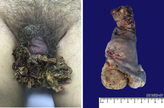
Figure 2 :
Macroscopic histopathological cuts with neoplastic invasion of both corpora cavernosa.
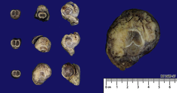
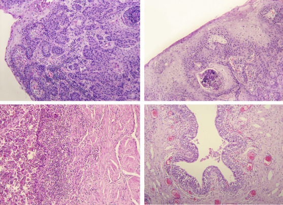
Figure 3 :
A. Infiltrating squamous cell carcinoma with high mitotic activity. B. Ulcerated and abscessed epidermoid cancer. C. Tumor infiltration to the corpus cavernosum. D. Urothelium without tumoral involvement.