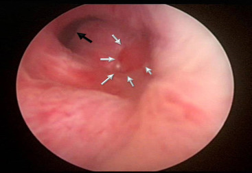
Figure 1 :
Urethroscopy at 6weeks: a 3×4cm non-epithelialized area in the realignment gap lined by connective tissue (white arrows). The black arrow points to the prostatic urethra.
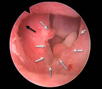
Figure 2 :
Urethroscopy at 12weeks (same patient): most of the non-epithelialized area has been covered by mucosa leaving a 1×0.5cm area where the deeper connective tissue layer was still exposed (white arrows). The Foley catheter in this patient was removed at week 15 when epithelialization was complete. The black arrow points to the verumontanum and prostatic urethra.